
Submitted by jer5150 on Thu, 08/16/2012 - 20:01
INTERPRETATION:
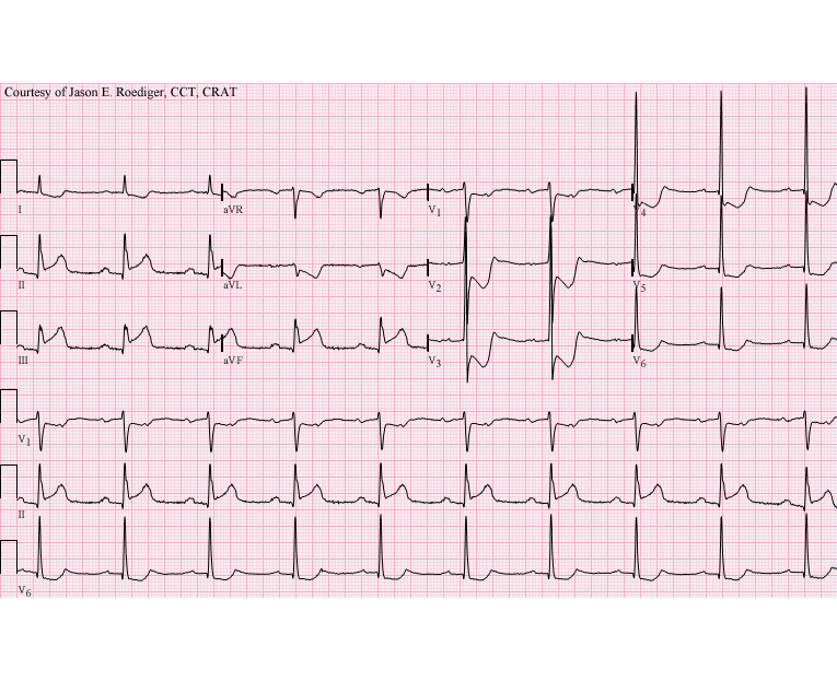
(1.) Sinus tachycardia (rate about 114/min) with . . .
(2.) . . . acute inferoposterior infarction complicated by . . .
(3.) . . . 2:1 Type I AV (nodal) block reducing the ventricular rhythm to a bradycardic rate of about 57/min.
COMMENTS: Take note of the blocked sinus impulses in monitoring lead V1 (3rd line up from the bottom of the ECG) that are superimposed on the apex of the inverted T-waves. The P-waves march out absolutely, precisely regular from left to right. That level of subtle detail doesn't reveal itself in any of the other 11 leads where the sinus impulses blend seamlessly into the contours of the T-waves. Without the benefit of V1, it would be impossible to know that the sinus rate (i.e., 114/min) is exactly twice that of the ventricular rate (i.e., 57/min). I don't know for sure but, given the circumstances, I wouldn't be surprised if this patient had recently received some atropine. Over the years I've repeatedly noticed that healthcare providers have a hard time understanding the concept of how a tachycardia can somehow result in a bradycardia but it makes sense if the ratio is 2:1. Tell someone that there is a "tachycardia" going on in this ECG and they look at you funny because all they are focused on is the rate of the QRS complexes. Since the computer failed to recognize the 2:1 ratio, it was fooled into believing that this was sinus bradycardia with so-called "first-degree" AV block. The cardiologist diagnosed this as Type II AV block but this is a classical manifestation of Type I AV block. The P-R interval on the conducted beats is prolonged at about 0.26s and there is no bundle-branch block (BBB). The anatomic level of the block is undoubtedly in the AV node and not infranodal. Just 2 1/2 hours prior to this ECG, the patient was in normal sinus rhythm and occasionally having associated periods of Wenckebach. In the following half-dozen years, the patient continued to experience Type I AV block with 3:2 Wenckebach periods and "escape-capture bigeminy". 7 years after their acute infarction, the patient eventually received an permanent pacemaker. Unbelievably, the cardiologist also diagnosed this as "Anterior myocardial infarction, possibly acute" when this is clearly acute inferoposterior infarction!
CLICK HERE ---> ---> ---> "DOWNLOAD ORIGINAL" <--- <--- <--- TO OPEN A NEW BROWSER WINDOW WHERE YOU CAN VIEW AN ANIMATED "GIF" OF 3 IMAGES THAT CONTINUOUSLY LOOP AT A TIME INTERVAL OF EVERY 10 SECONDS. THE INVERTED AND "EXPANDED" VIEW OF THE ECG ALLOWS YOU TO BETTER VISUALIZE THE INVOLVEMENT AND EXTENSION INTO THE POSTERIOR WALL WITHOUT OVERLAPPING OF THE WAVEFORMS IN THE ANTEROSEPTAL LEADS. IT'S EASIER THAN HOLDING UP AN INVERTED HARDCOPY IN FRONT OF A BATHROOM MIRROR!
Rate this content:
-

- jer5150's blog
- Log in or register to post comments
All our content is FREE & COPYRIGHT FREE for non-commercial use
Please be courteous and leave any watermark or author attribution on content you reproduce.