
Submitted by Dawn on Mon, 03/18/2013 - 14:10
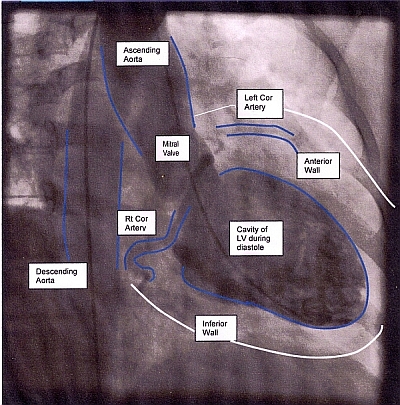
The ECG Guru now has a You Tube site where you can find videos to enhance your classes. As with all ECG Guru content, there is no charge and no copyright. Ventriculograms are often obtained during a cardiac cath procedure. A curved, or pigtail, catheter is inserted through the arterial access line into the aorta and then the left ventricle. Contrast is introduced into the left ventricle and the pumping function of the ventricle can be observed. The structure and function of the aorta and mitral valve may also be observed during this procedure. Unless you and your students are already accustomed to looking at ventriculograms, they may seem confusing, but they offer a priceless look at the working left ventricle. I am including here a labelled still photo of an LV gram, catching the LV in diastole. It is seen from the side, with the anterior wall above, and the inferior wall below. The muscle walls don't stand out on the xray, but in the moving version, their action can clearly be seen as the cavity of the LV squeezes. Watch our YouTube channel for ventriculograms illustrating poor ejection fraction, alcoholic myopathy, mitral regurgitation, and many other interesting things!
We currently have posted a great case study showing the effects of delay to treatment of M.I. If you show your students a normal ventriculogram, then this one showing akinesis of the inferior wall, they will clearly understand the stunning effects of M.I. on the heart muscle. Remind them that these effects are sometimes temporary, but can be permanent!
We put a larger version of this illustration on our "Resources" page.
Rate this content:
-

- Dawn's blog
- Log in or register to post comments
All our content is FREE & COPYRIGHT FREE for non-commercial use
Please be courteous and leave any watermark or author attribution on content you reproduce.