
Submitted by Dawn on Mon, 11/28/2011 - 18:57
We caution students that the signs of acute M.I. (ST elevation) cannot reliably be seen in cases of wide QRS. This is because, in wide QRS situations like left bundle branch block, ventricular rhythms, or right ventricular pacing, the ST segments will elevate in leads with downward QRS complexes, and depress when the QRS is upright. These is called discordant ST changes.
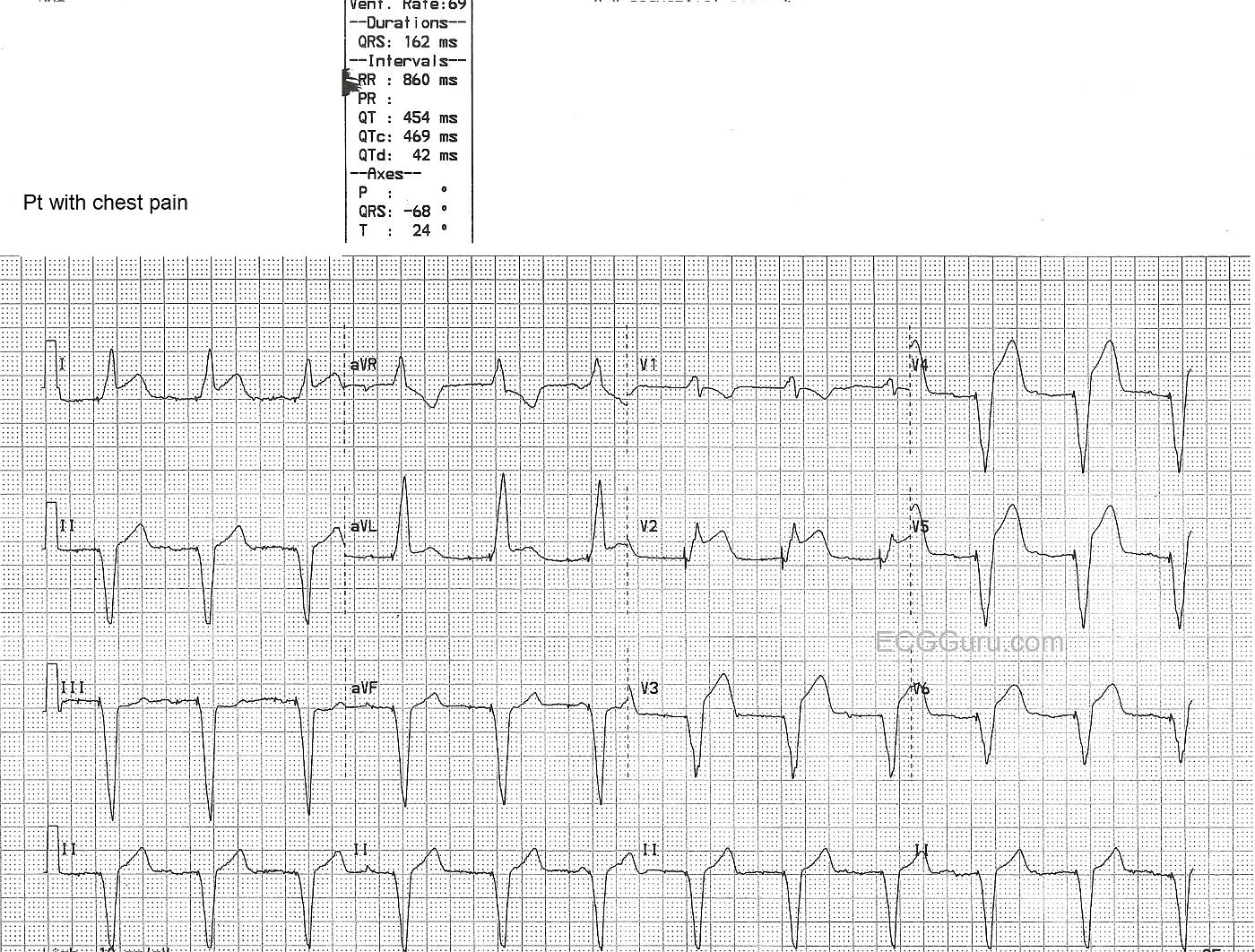
In this ECG, a man in his 60's presented with chest pain. His ECG showed AV sequential pacing, with ventricular pacing from the right ventricle. The QRS is 162 ms in duration. He has ST segment elevation in Leads I, aVL, and Leads V2 through V6.
The ST elevations are more pronounced than expected in this paced patient. But, the real clue here is the ST elevation in Leads I, aVL, and V2 - leads that should have ST depression because of their upright QRS complexes, have elevation! This patient was taken to the cath lab and the left coronary artery wass reperfused and stented. For more information about ST elevation in wide QRS complex rhythms, see this LINK.
Related Terms:
Rate this content:
All our content is FREE & COPYRIGHT FREE for non-commercial use
Please be courteous and leave any watermark or author attribution on content you reproduce.