Displaying 1 - 3 of 3
Dawn's picture

Ask The Expert

Dr. Jerry W. Jones, MD, FACEP, FAAEM has graciously shared with us his four-part article on the topic of “Delays & Blocks Involving the Bundle Branches”.

Dr. Jones is a talented instructor who makes difficult topics easy.  Please feel free to post your comments and questions for Dr. Jones and our other ECG Gurus. 

Click THIS LINK for a downloadable pdf of Part 1: Non-Specific Intraventricular Conduction Delays. 

Click THIS LINK for a downloadable pdf of Part 2: Left Bundle Branch Block.

Click THIS LINK for a downloadable pdf of Part 3: Right Bundle Branch Block.

Click THIS LINK for a downloadable pdf of Part 4: The Fascicles of the Left Bundle Branch 

Dawn's picture

Tachycardia In An Unresponsive Patient

 The Patient     This ECG was obtained from a 28-year-old woman who was found in her home, unresponsive.  She was hypotensive at 99/35.  No one was available to provide information about past medical history or the onset of this event.

Before you read my comments, pause to look at the ECG and see what YOU think.  We would welcome comments below from all our members!

The ECG     This ECG is quite challenging, as it illustrates the helpfulness of ECG changes in patient diagnosis, and also points out how important clinical correlation is when the ECG suggests multiple different problems. Forgive me in advance, but there is a lot to say about this ECG.

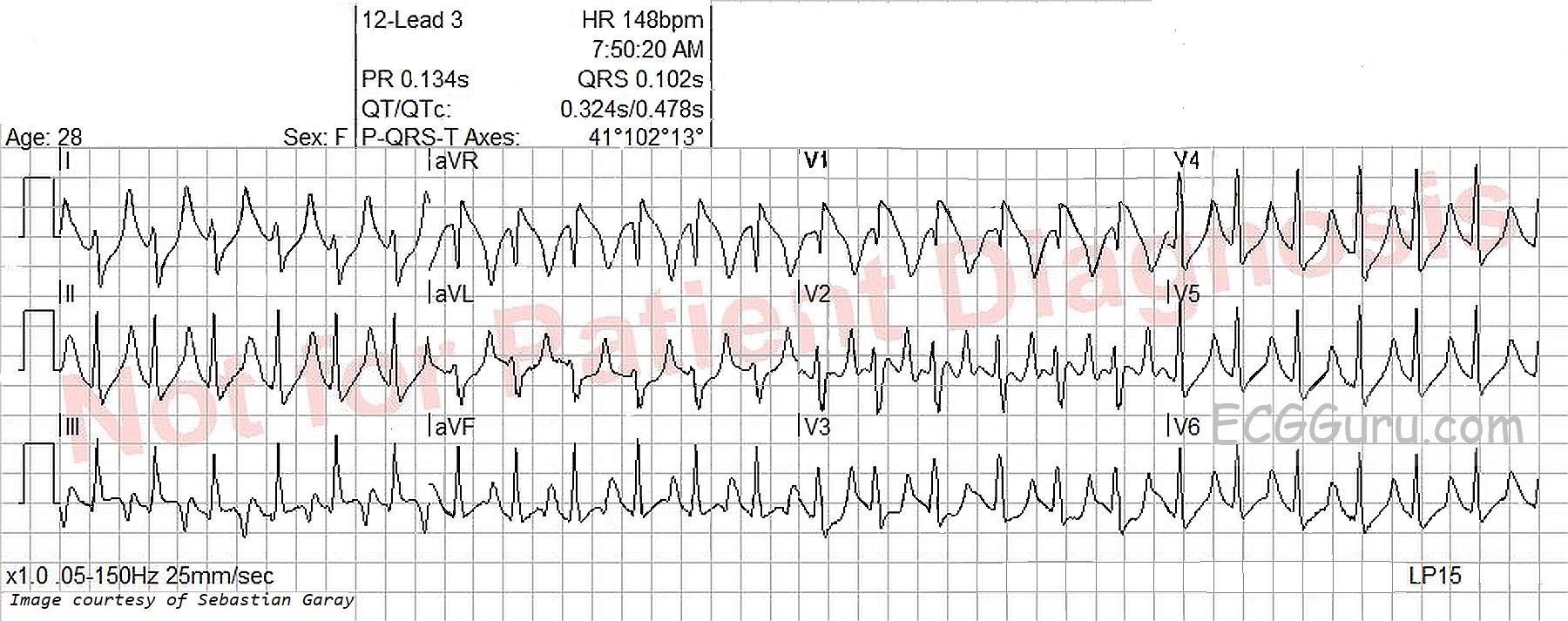
The heart rate is 148 bpm, and the rhythm is regular, although not perfectly. P waves are not seen, even though the ECG machine gives a P wave axis and PR interval measurement. The rate is fast enough to bury the P waves in the preceding T waves, especially if there is first-degree AV block. Differential dx: sinus tachycardia, PSVT, atrial flutter. The very slight irregularity points more towards sinus tachycardia.  The rate of nearly 150 suggests atrial flutter with 2:1 conduction, but the only lead that looks remotely like it has flutter waves is V2. The lack of an onset or offset of the rhythm makes it difficult to diagnose PSVT with any certainty.

Dawn's picture

Wide QRS Complex With First-degree AV Block

The Patient:  This ECG was taken from a 73-year-old man with a history of heart failure with preserved ejection fraction, severe left ventricular hypertrophy, Type II diabetes, and stage 4 chronic kidney disease.  He also suffered deep vein thrombosis and is on anticoagulation.  He has a recent diagnosis of IgA myeloma.  He presented with a complaint of nausea and vomiting and was found to have a worsening of acute kidney infection.  There was suspicion of renal and cardiac amyloidosis, but the patient refused biopsy to confirm this.  He was started on chemotherapy for multiple myeloma and will be followed as an outpatient.

The ECG:  The rhythm is sinus at around 60 bpm, although the rate varies a little at the beginning of the strip.  The QRS complex is wide at .12 seconds, or 120 ms., representing interventricular conduction delay (IVCD).  The PR interval is .32 seconds, or 320 ms. This constitutes first-degree AV block.  There is left axis deviation in the frontal plane and poor R wave progression in the horizontal plane.

All our content is FREE & COPYRIGHT FREE for non-commercial use

Please be courteous and leave any watermark or author attribution on content you reproduce.