Dawn's picture

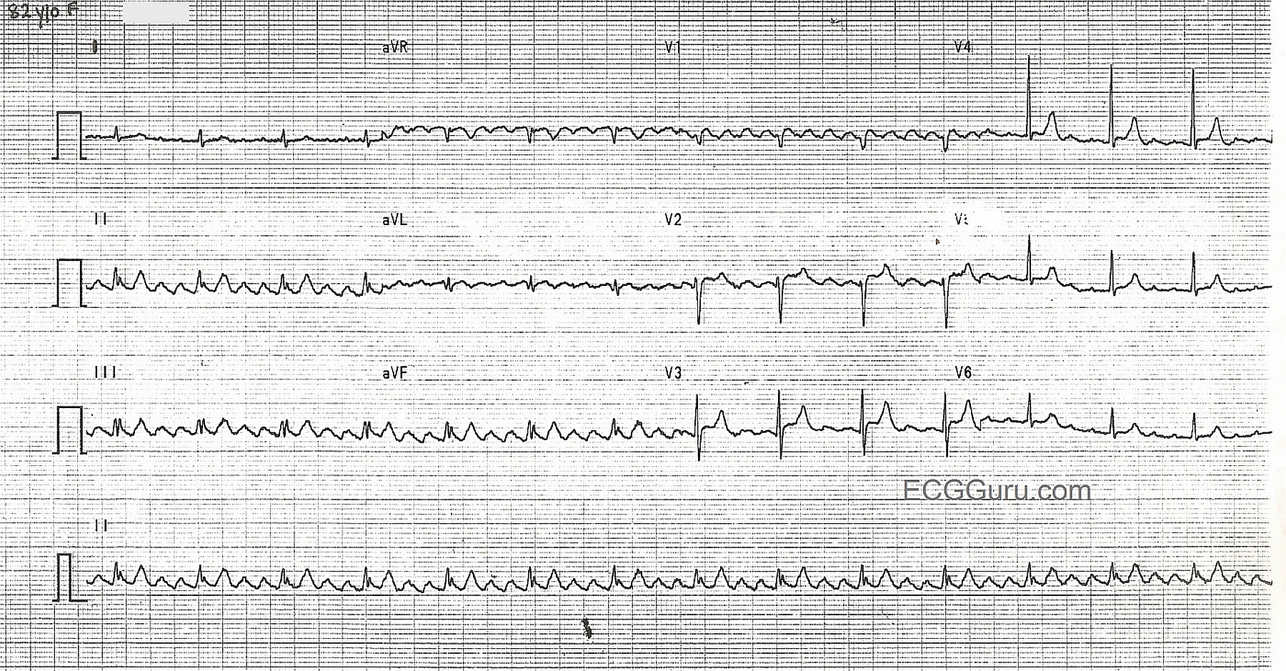
This nice example of atrial flutter offers many teaching opportunities. "Sawtooth" flutter waves are readily visible in Leads II, III, avF, and V1, typically good "P wave" leads.  Other leads show small, discreet P waves that the beginning student might not recognize as flutter waves because they don't produce the sawtooth pattern.  The P waves' rate, approximately 360/min., gives them away as atrial flutter.  This is also a good example of a constant 4:1 conduction, resulting in a regular QRS rhythm and regular pulse, at a rate of about 90/min.  This demonstrates that not all narrow-complex rhythms between 60 and 100 bpm are "NSR".  The low voltage in the limb leads makes this a good ECG to demonstrate that the flutter waves are regular and do not pause when the QRS happens, illustrating the separate actions of the atria and the ventricles.  For students just learning 12-lead interpretation, this ECG serves to show that acute ST elevation M.I. is not the only valuable information that can be obtained from a 12-Lead, and that some leads are better than others for showing dysrhythmias.

Rate this content: 
Average: 4.7 (3 votes)

Comments

ekgpress@mac.com's picture

Dawn's January 19, 2013 Instructor Collection ECG is of Atrial Flutter with 4:1 AV Conduction. LOTS of important teaching concepts are conveyed by this tracing. These include:

  • It is well to keep in mind that AFlutter is the most commonly misdiagnosed/overlooked sustained arrhythmia. While large amplitude flutter waves in this tracing pose no difficulty to diagnose - it is easy to imagine how if only lead I was used, that the diagnosis of AFlutter would not be apparent. Often - there are only a few leads that show flutter, emphasizing again the concept that "12 leads are better than one" for arrhythmia diagnosis.
  • BEYOND-the-CORE: With typical AFlutter (which makes up >80% of cases) - the sawtooth pattern of flutter is extremely well visualized in the inferior leads and in lead V1. The flutter wavefront almost always manifests CCW (counterclockwise) rotation around the tricuspid ring. Occasionally - typical AFlutter will follow the same path through the atria, but with CW (clockwise) rotation around the tricuspid ring (ie, "reverse" typical flutter). In such cases - the same leads reveal the diagnosis, but instead of upright flutter waves in V1 there will often be a double negative (W-shaped) flutter wave in V1. While NOT important for non-electrophysiologists to distinguish between these forms of AFlutter on the surface ECG - it IS clinically relevant to be aware that atypical AFlutter forms may be seen in 10-20% of cases, in which the path through the atria is different and AFlutter may be present despite absence of a prominent "sawtooth" pattern. As a result - atypical AFlutter may be more difficult to recognize on ECG ... Treatment considerations (ie, referral to EP with consideration for ablation) remain similar. For an excellent PDF review by Garcia-Cosio (including color pictures of the various forms and rotations of AFlutter) - CLICK HERE - Be sure to check out Figure 2 on page 818 in this pdf.
  • The other clinicallly relevant point raised in my last bullet is that what used to be classified as "atrial tachycardia" in non-digoxin-toxic patients is now often referred to as "atypical" AFlutter.
  • BACK-to-BASIC AFLUTTER ECG CONCEPTS: AFlutter is characterized by a special pattern of regular atrial activity that in adults almost always (and almost magically) occurs at a rate of ~300/minute (250-to-350/minute = usual range). The most common ventricular response to AFlutter (by far! ) - is with 2:1 AV conduction. As a result, the ventricular rate with untreated AFlutter will usually be close to 150/minute (ie, 300/2). NOTE: The atrial rate may be less than 250-300/minute - IF the patient is already taking certain antiarrhythmic drugs.
  • The next most common AV conduction ratio with AFlutter is 4:1 AV conduction (atrial rate ~300/min; vent. rate ~75/minute). This is the situation for this tracing. The flutter rate is at the upper limit of the usual range - with only 1 out of every 4 flutter waves being conducted to the ventricles.
  • This tracing does not represent "AV block" - since conduction is physiologic. Bombardment of the AV node with 300-plus flutter impulses each minute overwhelms the AV node capacity to conduct - so only 1 out of 4 beats make it to the ventricles. NOTE that the F-R interval (distance between the last flutter wave in each cycle until the QRS) is constant! - so there IS conduction! Interestingly - it is never the closest flutter wave that conducts (concealed conductions prevents that).
  • Occasionally - AFlutter may manifest a variable (=irregular) ventricular response. In such cases - there will be an irregularly irregular ventricular response resembling AFib except for the presence of regularly occurring sawtooth flutter waves.
  • Odd conduction ratios (1:1; 3:1; 5:1) are uncommon for AFlutter.
  • Finally - Note how challenging it is to assess ST-T waves in this tracing due to ongoing distortion by AFlutter ...
  • For anyone with an interest - Please Check out my ACLS COMMENTS #9 on Narrow Tachycardias, that includes a case discussion SECTION on AFLUTTER.

Ken Grauer, MD  www.kg-ekgpress.com   [email protected] 

All our content is FREE & COPYRIGHT FREE for non-commercial use

Please be courteous and leave any watermark or author attribution on content you reproduce.