Displaying 1 - 10 of 32
Dr A Röschl's picture

High-grade AV Block

Why is this a high-grade AV block? If at least 3 P-waves are not conduced and there is normal AV conduction before and after, this can be considered a high-grade AV block. In this Holter strip, P1, P2 and all P-waves from P6 onwards are conducted, albeit with a prolonged PR interval (first-degree AV block). P3, P4, P5 are not conducted. A junctional escape beat is seen before P5. P5 can also not be conducted because the specific conduction system is still refractory at this time due to the junctional escape beat.

Furthermore, a long QT time is observed!

Dawn's picture

Complete Heart Block or High Grade AVB?

The patient:  This ECG was obtained from a 91-year-old woman who was complaining of weakness.  Unfortunately, we have no other information. 

The ECG:  This ECG has something for your basic students, and even more for the more advanced learners.  The first thing  that anyone should notice is the slow rate.  The ventricular rate is around 35 bpm, and regular.  If the patient is showing signs of poor perfusion, we would stop here and prepare to increase the rate with a temporary pacemaker (transvenous or transcutaneous). Why is the rate so slow?  There is no P wave in front of each QRS, so this is not sinus bradycardia.  Rather, we see P waves at a rate of approximately 100 bpm, wit a very regular rhythm.  Beginners should “march out” the P waves with calipers or by marking a straight edge piece of paper.  There are 15 P waves on this ECG – some are buried within QRS complexes (QRS #3) or T waves (QRS #4).

 Because there are two distinct, regular rhythms, but they do not track with one another, we know this is possibly third-degree AV block (complete heart block).  Another clue is that there are no steady, repetitive PR intervals, which means there is no relationship between the atrial rhythm and the ventricular rhythm.

 For more advanced learners, it is helpful to try to identify the origin of the escape rhythm.  If it is junctional, the AV block is above the junction.  If the escape is ventricular, the AV block is below the junction.  A junctional rhythm is usually between 40 – 60 bpm, with a narrow QRS.  Ventricular escape rhythms are usually less than 40 bpm and with wide QRS complexes.   This ECG will be a little challenging on this front, because the rhythm has some characteristics of junctional rhythm and of ventricular rhythm.

Dr A Röschl's picture

HIGH GRADE AV-Block

Dr A Röschl's picture

Why is this not second degree AVB Type II and no high grade AVB

(Image 1) Why is there no second-degree AVB  Mobitz type II and no high-grade AV block? To the first question: Basically, second-degree AV block Mobitz type II is rare. The two ECG patterns that can easily be confused with Type II Mobitz block are: blocked/non-conducted PACs and second-degree AVB Mobitz type I (Wenckebach). (Image 2) You have to compare the PR duration before the pause and after it. With the naked eye, the difference is often difficult to recognize, a pair of calipers does a good job here.

Dr A Röschl's picture

Second degree AVB Mobitz Type II

This ECG is from an 80-year-old lady who has collapsed or had sycopal episodes several times. The ECG comes from a Holter monitor. She has arterial hypertension and coronary artery disease. The ECG shows a second-degree, Mobitz Type II AV block. In both types of AVB, the PP intervals are usually the same.

Dawn's picture

ECG Basics: Second-degree AV Block, Type I

This two-lead rhythm strip shows a normal sinus rhythm at about 63 bpm.  The P waves are regular. After the sixth P-QRS, there is a non-conducted P wave.  The normal rhythm then resumes.  The two most common reasons for a non-conducted P wave in the midst of a normal sinus rhythm are 1) non-conducted PAC, and 2) Wenckebach conduction. The first is easy to rule out.  The non-conducted P wave is not premature, so it is not a PAC.  The second one is a little harder when we only have a short strip to look at.  We are conditioned to look for progressively-prolonging PR intervals until a QRS is "dropped".  In this case, the progression is in very tiny increments that are hard to see unless you zoom in and measure.  But they ARE progressively prolonging.  An easy hack:  measure the last PRI before the dropped beat and the first one after the pause.  You will see that the cycle ends on a longer PRI (about .28 seconds) and the new cycle starts up with a PR interval of about .20 seconds.  Fortunately, this conduction ratio will have very little effect on the patient's heart rate.

Dawn's picture

ECG Basics: Second-degree AV Block, Type II

This rhythm strip was obtained from a man who was suffering an acute inferior wall M.I.  There are ST elevation and hyperacute T waves.  The rhythm is SINUS ARRHYTHMIA WITH SECOND-DEGREE AV BLOCK, TYPE II.    There is also first-degree AV block.

There are more P waves than QRS complexes, with a 3:2 ratio.  The atrial rate varies between 55 -68 beats per minute.  The sinus rate speeds slightly after the dropped QRS in each group. The ventricular rate is about 40 bpm, with grouped beating. (Regularly irregular.)

The PR intervals are steady at 226 ms (slightly prolonged).

Dawn's picture

Second-degree AV Block, Type II

The patient:  Unfortunately, we no longer have information on this patient, other than the fact that she went to the OR for a permanent pacemaker implantation.

The ECG:  The atrial rate (P waves) is 99 beats per minute. The P waves are regular and all alike (NSR). The ventricular rate (QRS complexes) is 33 bpm, and the QRS complexes are regular and all alike. The PR intervals, when A-V conduction occurs, are 162 ms (.16 seconds) and all alike. The QRS complexes are wide, at 122 ms (.12 seconds). There is right bundle branch block, but no left hemiblock, as the frontal plane axis is normal. The QTc is prolonged at 549 ms.  Many ST segments on this ECG have a “flat” appearance, rather than the normal concave up shape.

The failure of 2 out of every 3 P waves to conduct indicates a second-degree AV block. Type I is a block of the AV node, with progressive prolongation of the PR interval until ONE P wave fails to conduct. Type II AV block is a block of the intraventricular conduction system. Clues that a second-degree AV block is Type II include:

·        The PR intervals are all alike.

·        More than one consecutive P wave is not conducted.

·        A P wave that is NOT in the refractory period of the preceding beat is not conducted.

Dawn's picture

ECG Basics: 2:1 AV Block

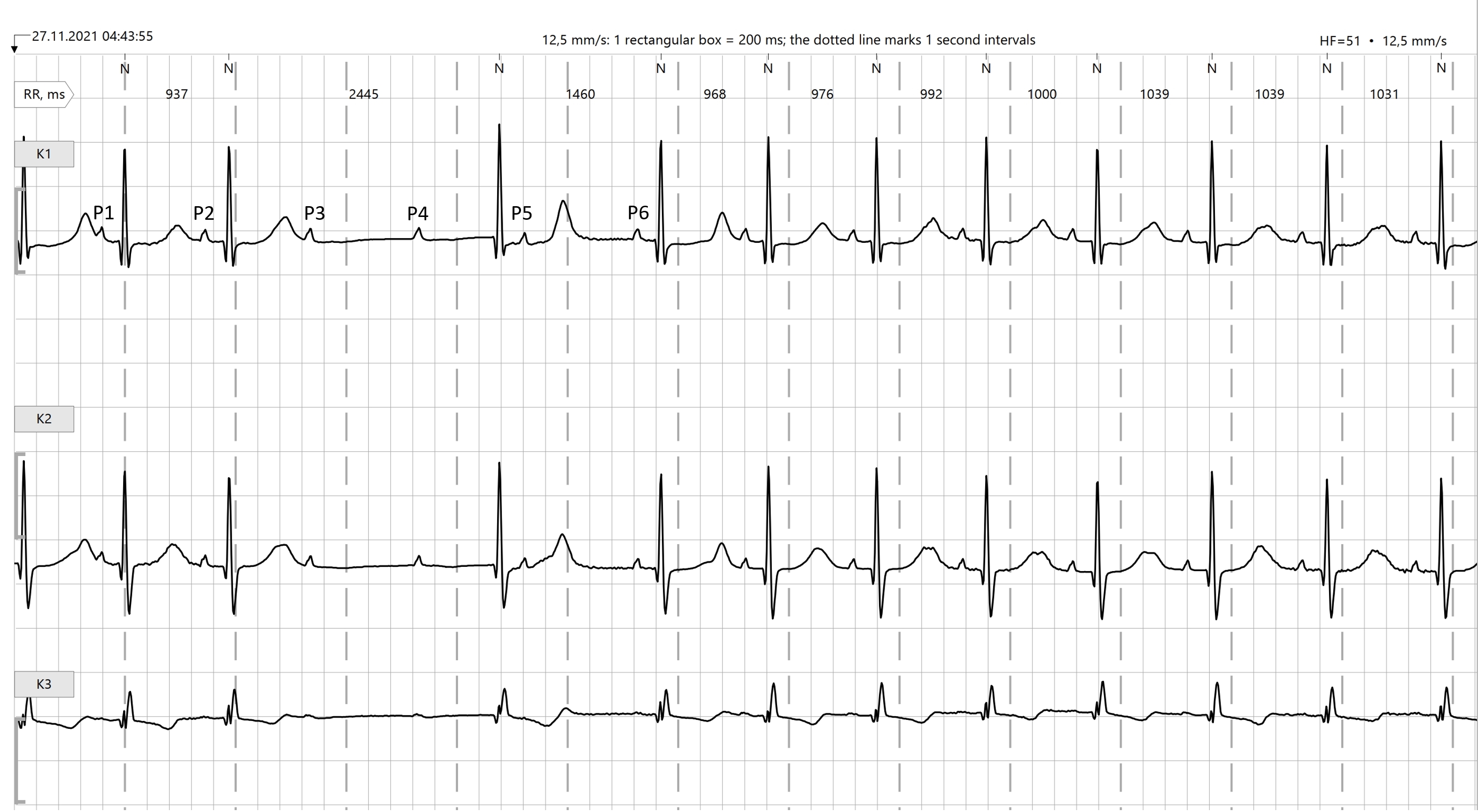
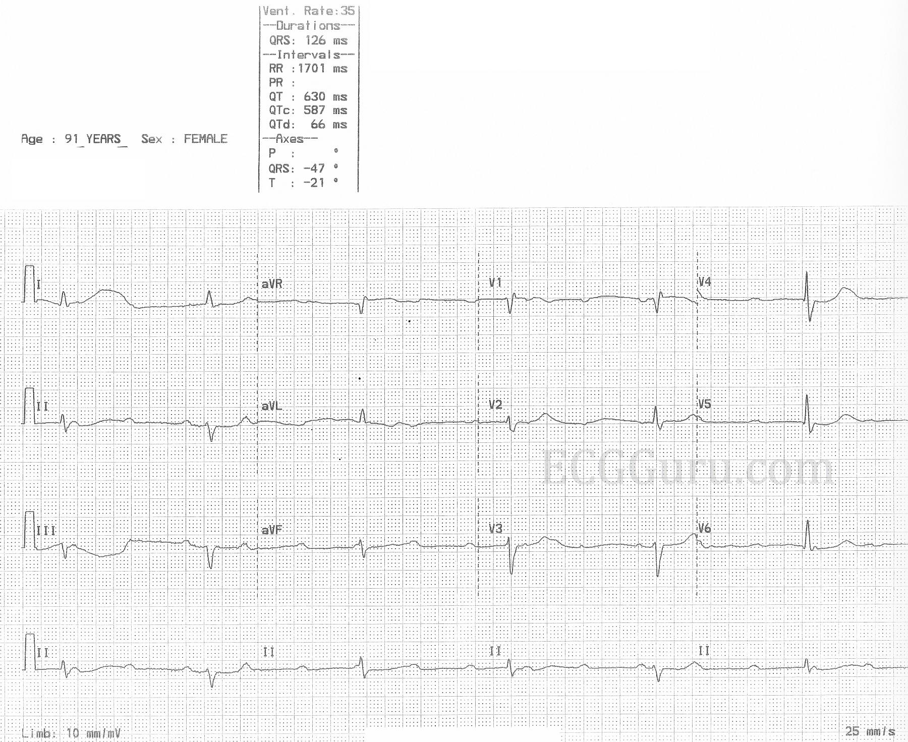
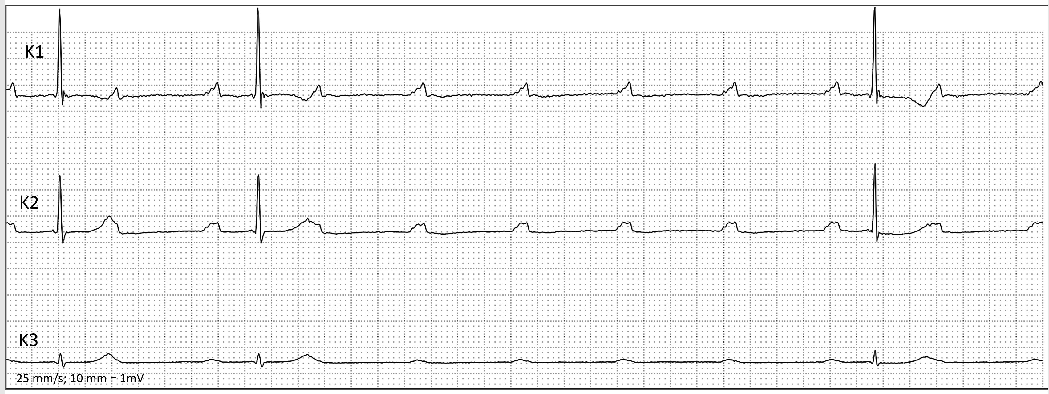
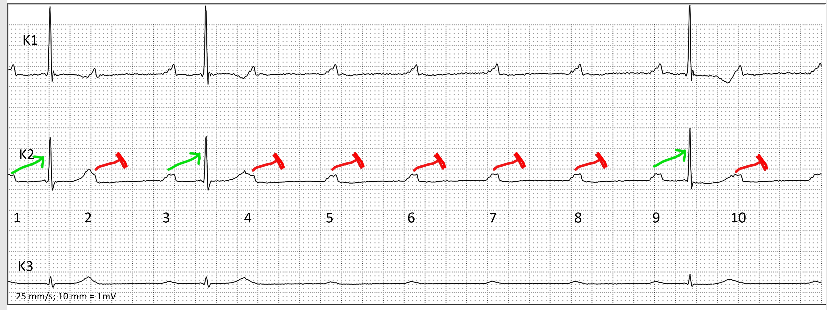
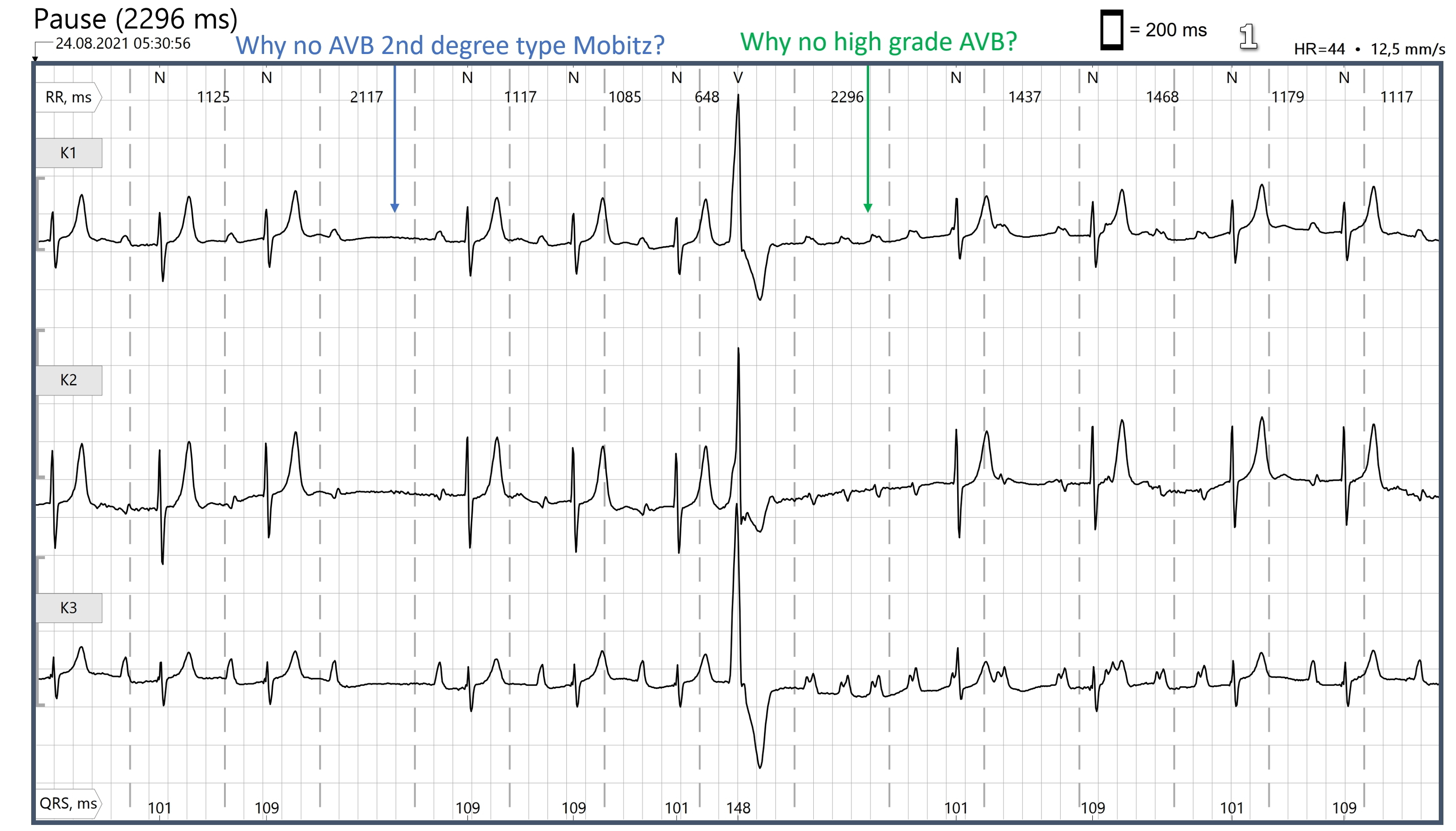
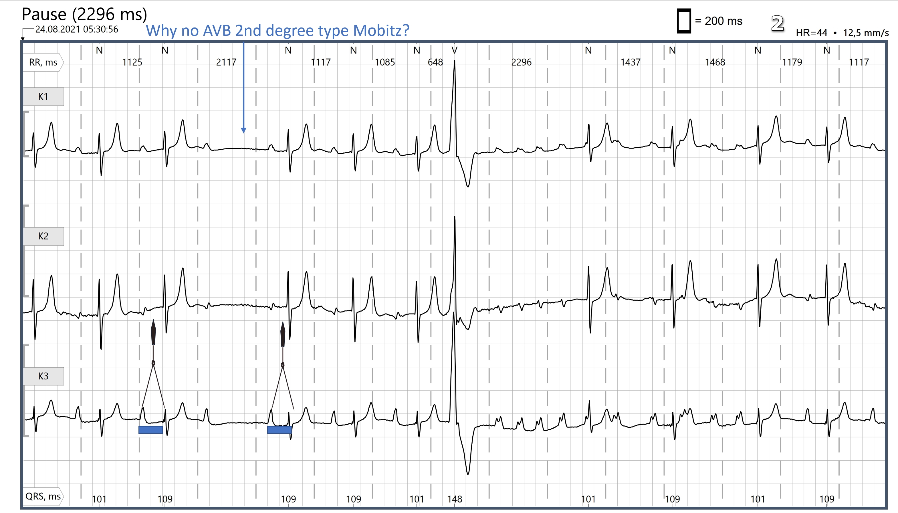
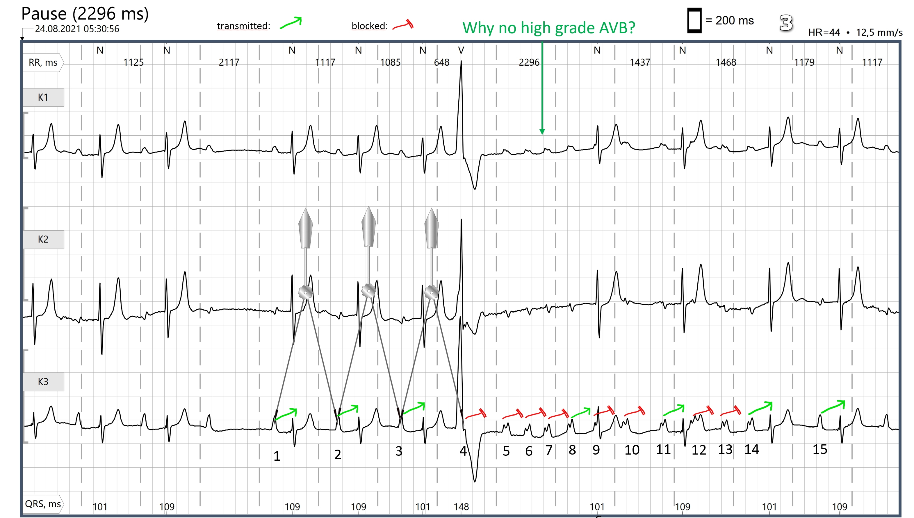
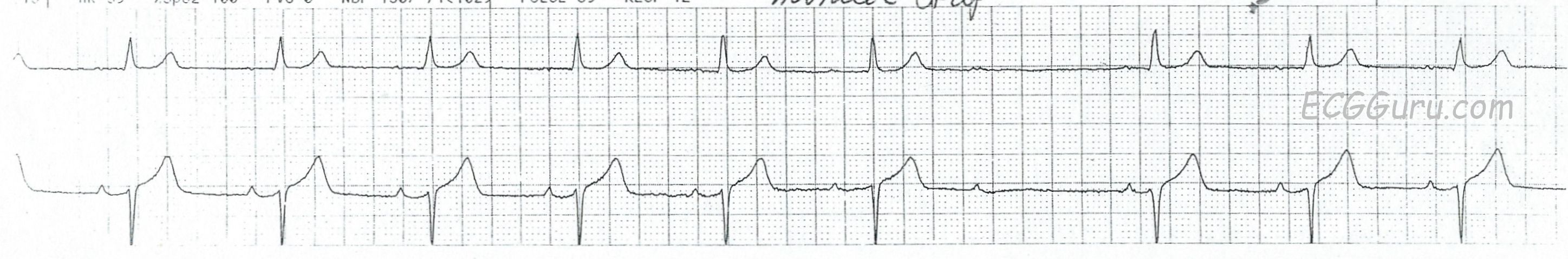
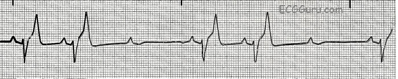
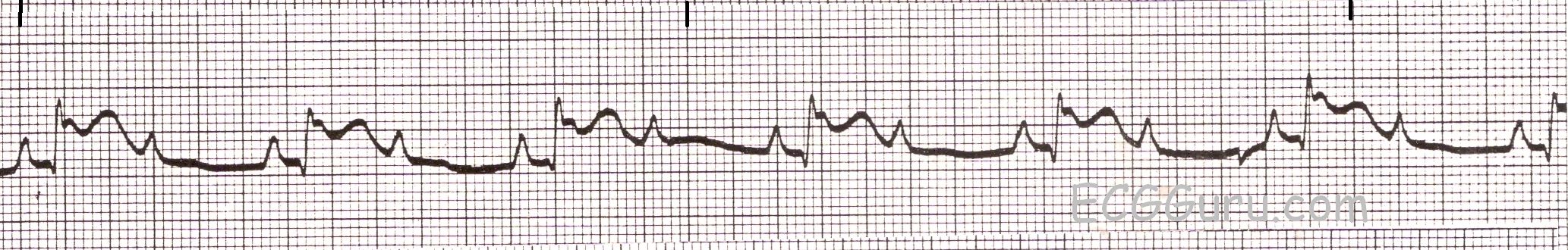
Second-degree AV block can either be Type I (Wenckebach) or Type II.  In either case, some P waves are conducted to the ventricles, and some are not. Type I blocks usually occur in the AV node, and are often benign. Type II blocks are more often "sub-Hisian", or fascicular blocks, and are more likely to progress to higher levels of AV block and bradycardia.  When a second-degree AVB is conducted in a 2:1 ratio, it is difficult to differentiate Type I from Type II.  Features that favor the diagnosis of Type I are narrow QRS complex and the non-conducted P waves land on the previous T waves - during the refractory period of the ventricles.

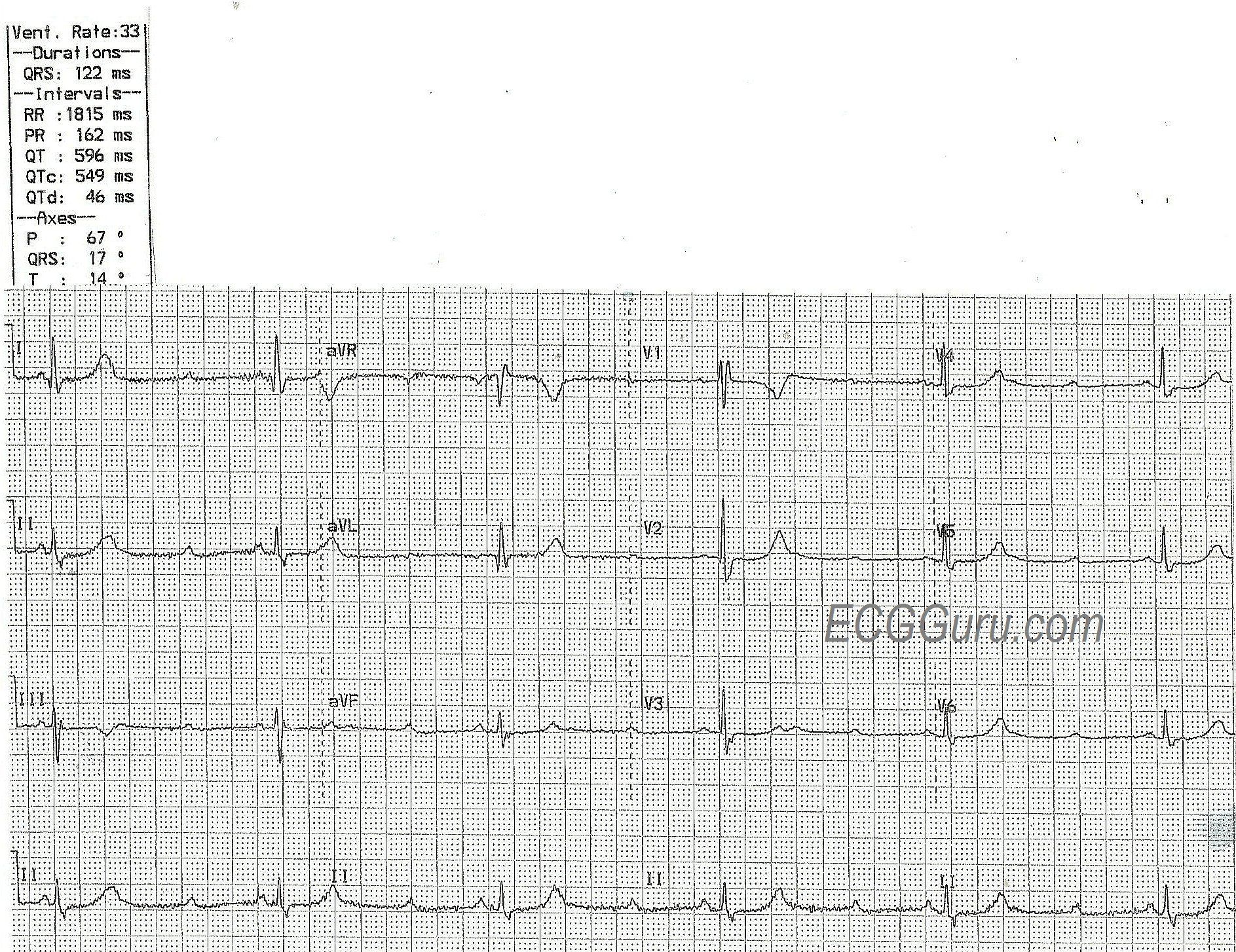
Type II blocks are more likely to have a wide QRS with a bundle branch block morphology.  That is because Type II blocks often reflect serious fascicular disease.  A typical Type II block is a persistent bifascicular block (ex: RBBB and left anterior hemiblock)) with an intermittent block in the third fascicle.  Another way to think of it is an intermittent tri-fascicular block. If that one remaining fascicle stops conducting, the patient will be in complete heart block.

Signs of Type II blocks include the wide QRS and also two or more non-conducted P waves in a row.  Also, P waves that are "out in the open", away from the refractory period, but fail to conduct are an ominous sign.

One strategy for reacting to a 2:1 block is to first assess the ventricular rate (54 bpm in this example).  Determine if it is adequate for the patient's hemodynamic stability.  If not, act to increase the rate.  Otherwise, it may be prudent in the stable patient to watch the rhythm strips for a while.  Sometimes, two p waves in a row will conduct - unmasking either progressive prolongation of the PR interval (Type I) or stable PR intervals (Type II). 

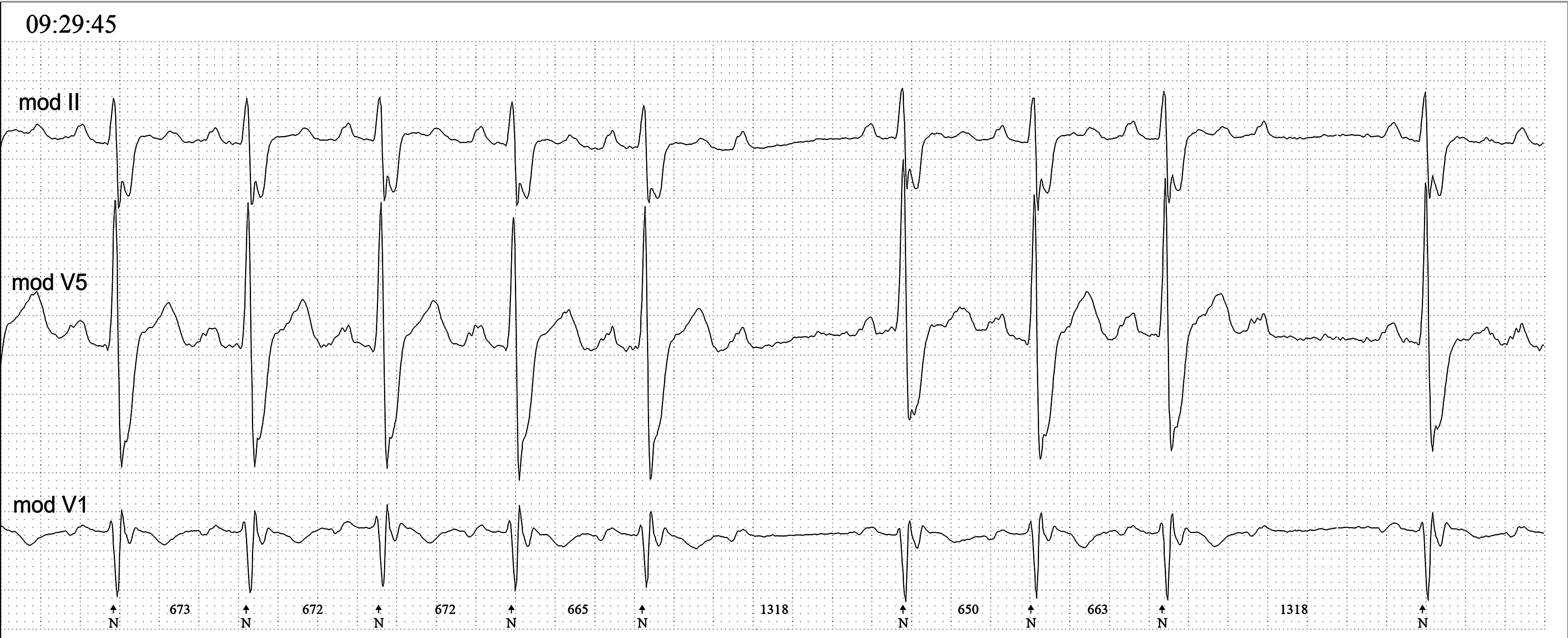
The patient in this example was having an inferior wall M.I.  The ST elevation will not always show up on a monitor strip, as it does here.  A 12-lead ECG is the minimum standard for evaluating for coronary artery disease and acute M.I.  It is possible that the 2:1 block will disappear when the atrial rate (about 108 here) is slowed.

Two Types of Complete Heart Block

Click to open: 
Two Types of Complete Heart Block

This is an original illustration by Dawn Altman. You may use it free of charge to enhance your presentations or student handouts. Click on image, or right click and SAVE image. For permission and charges for use in publications or for marketing uses, please contact the artist at [email protected]  

Pages

All our content is FREE & COPYRIGHT FREE for non-commercial use

Please be courteous and leave any watermark or author attribution on content you reproduce.