Displaying 1 - 10 of 11
Dawn's picture

Pediatric ECG: One month old infant

The patient:    4 week old female infant with past medical history of meconium aspiration at birth with APGAR scores of 2,4,6. Intubated and given nitric oxide for pulmonary hypertension. Weaned in NICU over 10 days. Echocardiogram during that time showed stiff pulmonic valve. This ECG was obtained at follow up appointment. Infant is eating well, no cyanotic spells. Four- week echo continues to show pulmonic valve stenosis. We do not know chamber measurements. Patient scheduled for a balloon valvuloplasty.  

Babies are born with relatively symmetrical hearts.  Unlike adult hearts, the right ventricle comparatively large due to the work it has to do to pump against the high pulmonary pressure before birth.  Because newborns’ hearts are more to the right side, we often do right-sided chest leads in addition to the standard 12-lead ECG.

 The ECG:   In spite of the pulmonary valve stenosis, this ECG is within normal limits for this 4-week old. Some of the ECG findings here that would be abnormal for an adult ECG, but are normal for this 4-week-old baby are: 

Dr A Röschl's picture

SINUS ARRHYTHMIA

Here we see the EKG of a 15-year-old girl. The rhythm is irregular, with the heart rate fluctuating between approximately 60 and 90 beats per minute. All P waves are identical, and the PR interval is always constant. Therefore, a sinus arrhythmia is present. In this case, it is a respiratory sinus arrhythmia, which is commonly found in younger individuals. The heart rate increases reflexively during inspiration and decreases during exspiration. This physiological sinus arrhythmia is usually no longer present in older individuals.

Dr A Röschl's picture

ECG OF A 3 YO BOY, IS IT NORMAL?

Here is the ECG of a 3-year-old boy. Is there cause for concern? The ECG shows a sinus rhythm with significant sinus arrhythmia. The heart rate increases with inspiration and decreases with expiration, which is called respiratory sinus arrhythmia. The QRS-axis is between 60 and 90 degrees, which is physiological at this age. The negative T-waves in V1-V3 (V4) are also age-appropriately normal. Therefore, there is no reason for concern; the ECG is considered normal for the child's age.

Dawn's picture

Narrow-complex Tachycardia In An Infant

The patient:  This ECG was obtained from a two-month-old girl who was a patient in the Emergency Department.  She had a fever due to a respiratory infection and was dehydrated. She was alert, active, and irritable.

The ECG:  There is a narrow-complex tachycardia at a rate of 194 bpm.  This is faster than the normal range for a two-month-old, which is about 80-160 bpm.  The intervals are all within normal range.  The frontal plane axis, at 145 degrees, is rightward, which is normal for this age. There are prominent, narrow Q waves in the inferior wall leads (II, III, and aVF) and in the left lateral leads (V4, V5, and V6).  There are no Q waves in the high lateral leads (I and aVL).  This is a normal pattern for this age group.   www.sciencedirect.com/science/article/pii/B9781416037743100280

The evaluation of this ECG must be preceded by a thorough evaluation of the patient.  SINUS TACHYCARDIA would be expected in the setting of fever, dehydration, hypoxia, pain or other discomfort. Should the rate fail to gradually return to a normal range after treatment, we would have to consider a reentrant supraventricular tachycardia. Reentrant tachycardias have a SUDDEN ONSET and SUDDEN TERMINATION.

Unfortunately, we do not have follow up on the patient.

Dawn's picture

Normal ECG in Seven-year-old Girl

The Patient:   This ECG was obtained from a seven-year-old girl who was complaining of mild, non-specific chest pain.  A medical workup found no structural or electrical abnormalities, and her pain resolved without treatment.  This is a good ECG to illustrate some of the ECG findings that are normal at this age, which might not be normal in adults or in infants.

The ECG: A general description of this ECG would include:

·         Normal sinus rhythm at 87 bpm

·         Normal frontal plane QRS axis at 60 degrees

·         PR interval:  .132 second (132 ms)

Dawn's picture

Supraventricular Tachycardia in Pediatric Patient With Wolff-Parkinson-White Syndrome

This ECG was obtained from a 13-year-old boy who had previously been in NSR.  At that time, he was diagnosed with Wolff-Parkinson-White syndrome, and he had delta waves.  He now presents with a supraventricular tachycardia.  People with W-P-W have one or more accessory pathways which allow atrial impulses to bypass the AV node and “pre-excite” the ventricles.  An accessory pathway, along with the normal pathway through the AV node, can form a functional circuit, allowing conduction to proceed down one pathway and return up the other, in a rapidly repeating circular motion.  This causes paroxysmal supraventricular tachycardia. 


In W-P-W, the most common type of SVT is AV reciprocating tachycardia (AVRT), also called AV reentry tachycardia.  Conduction through the accessory pathway can be forward (anterograde) or backward (retrograde).  The most common type of reentrant conduction in W-P-W is ORTHODROMIC, which we see here.  The impulse conducts forward through the AV node, and backward over the accessory pathway.  There is no delta wave, as there is no pre-excitation of the ventricles. 


Features of orthodromic AVRT are:

Fast rate (usually approximately 140-250 beats per minute)    

Narrow QRS complexes (unless BBB or IVCD present)   

P waves retrograde, if seen (negative in II, positive in aVR and V1)  

ST depression and T wave inversion common. 


Dawn's picture

ECG Basics: Retrograde P Waves

This Lead II rhythm strip shows a regular rhythm with narrow QRS complexes and retrograde P waves.  The strip was taken from a nine-year-old girl.  The rate is about 110 per minute and the PR interval is .12 seconds (120 ms).

When retrograde conduction is seen in the atria, it is often assumed that the rhythm is originating in the junction.  When a junctional pacemaker is initiating the rhythm, the atria and ventricles are depolarized almost simultaneously.  This can produce a P wave in front of the QRS with a short PR interval, during the QRS, or after the QRS.  Sometimes, in junctional rhythm, a block prevents the impulse from entering the atria, producing NO P wave.  Junctional rhythms are usually slow "escape" rhythms, but can be accelerated or tachycardic.

The fact that this rate is 110 / minute and the PR interval is normal at .12 seconds, we should consider that this rhythm could also be from an ectopic pacemaker low in the atria.  From this low starting point, the impulse will travel backward, in a "retrograde" fashion, through the atria, producing a negatively-deflected P wave in Lead II.

We do not have clinical data on this patient, and so do not know what possible causes of arrhythmia might be present, and what the expected rate should be in this situation.  

Dawn's picture

Pediatric ECG With Junctional Rhythm

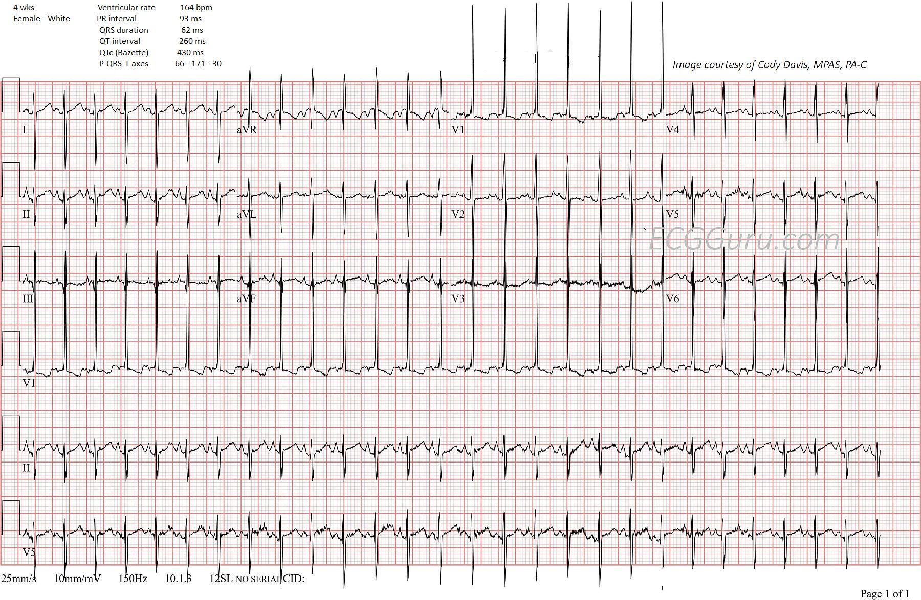
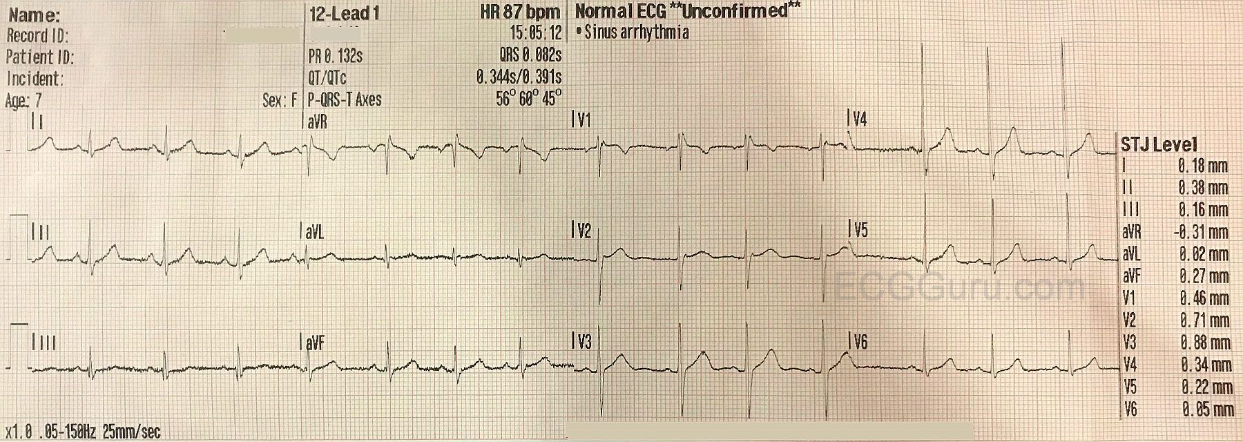
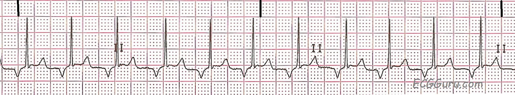
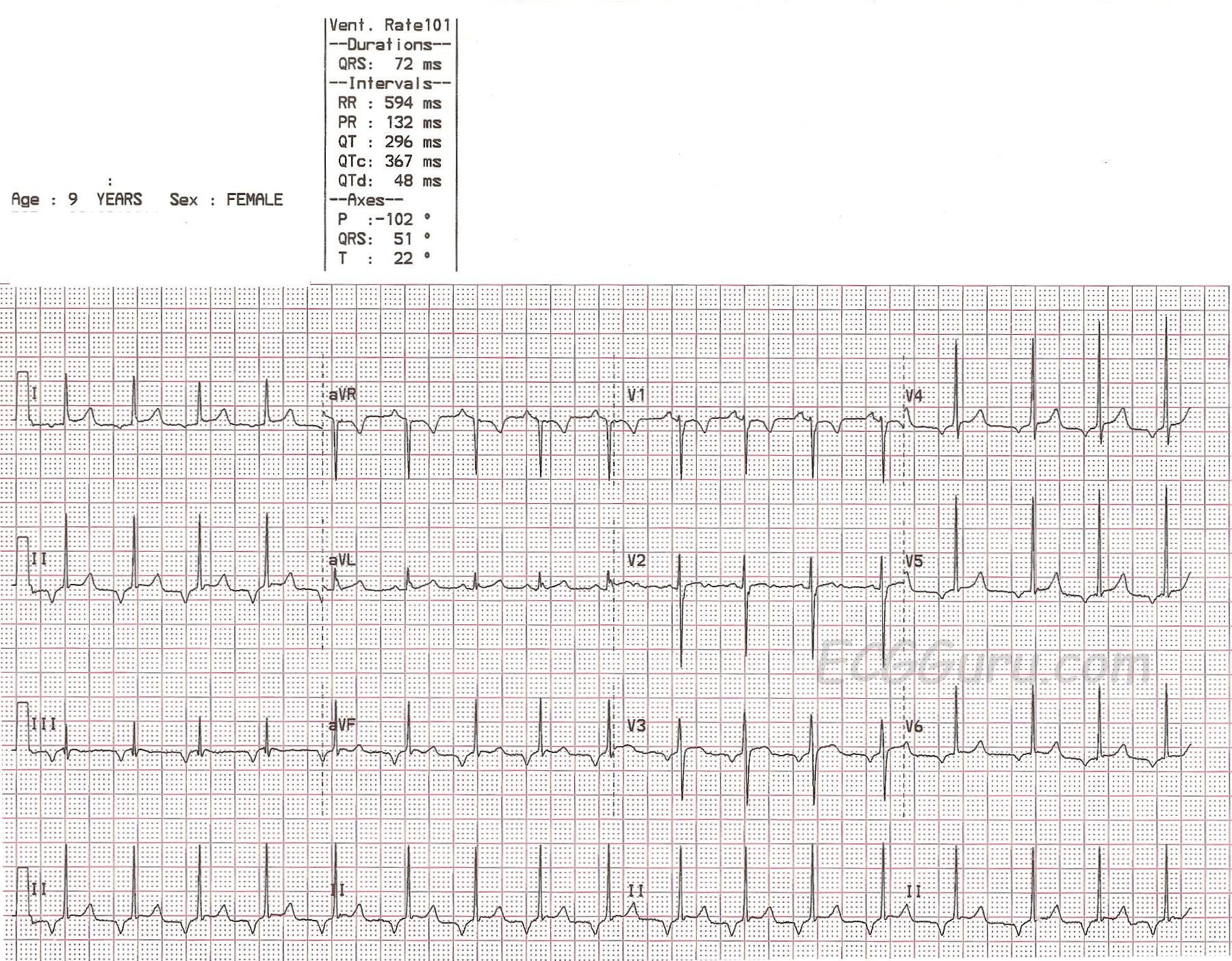
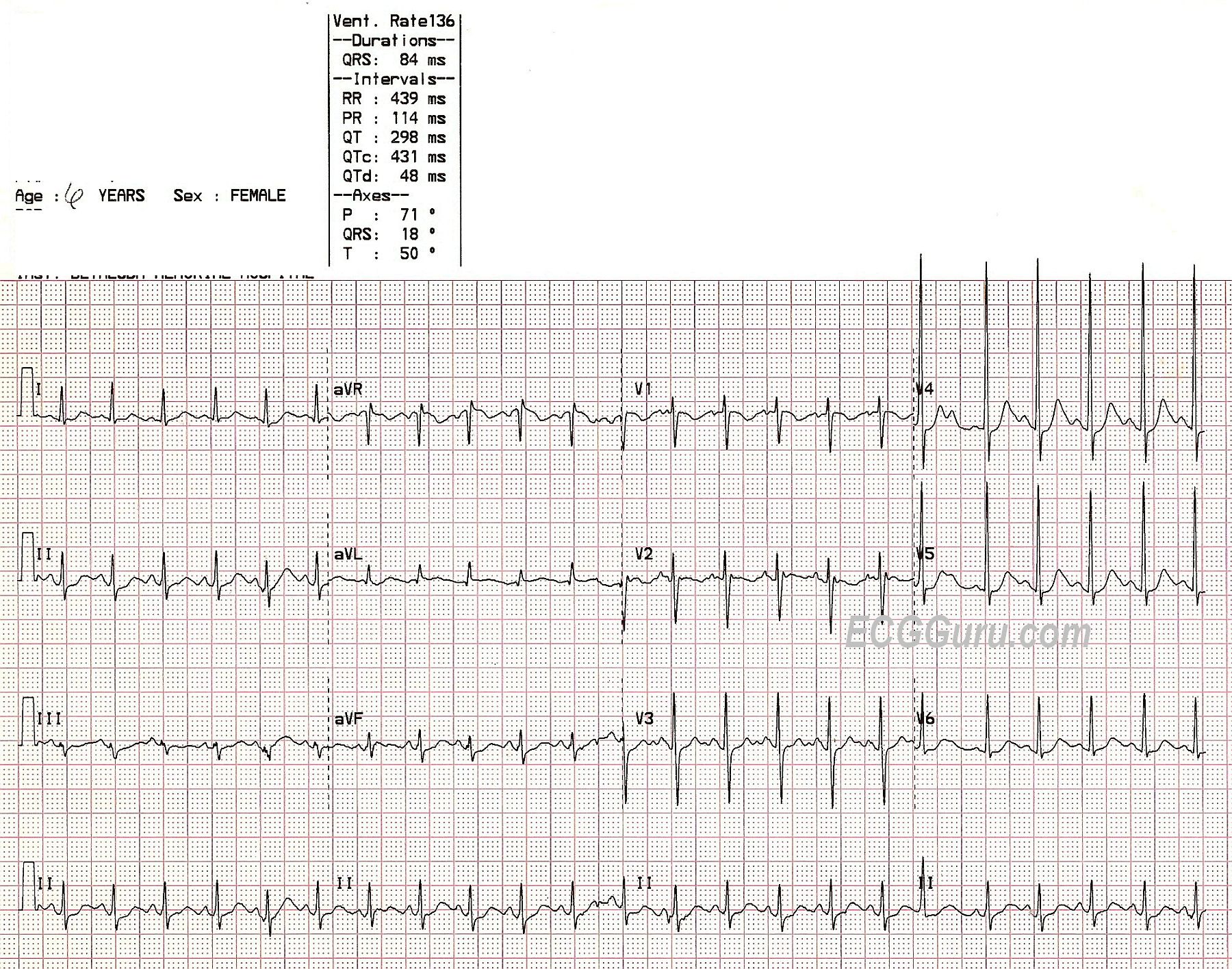
This ECG, taken from a nine-year-old girl, shows a regular rhythm with a narrow QRS and an unusual P wave axis. Unfortunately, we do not have any clinical information. 

Normally, P waves are positive in Leads I, II, and aVF and negative in aVR.  They can be biphasic in V1, but are usually positive in the rest of the precordial leads. . The P waves in this ECG are NEGATIVE in Leads I,II, III, aVF, and V3 through V6.  This indicates RETROGRADE conduction through the atria - the impulse starts low and continues in a backward fashion through the atria. This tells us that the rhythm originated in the AV junction or low atria.  The "junction" is usually defined as all of the complex AV node and the Bundle of His. 

The literature over the years has been very confusing about the exact location of the "junctional" pacemakers.  One commonly-accepted guideline was that a rhythm is "junctional" if there are retrograde P waves with a short PR interval, or a P wave that occurs within or after the QRS.  A rhythm with a retrograde P wave and a NORMAL PR interval is said to be "low atrial", indicating that the ectopic pacemaker involved was located in the low atrium, producing retrograde conduction through the atria and normal delay through the AV node.

While both of these scenarios are plausible, it probably is not possible to say with certainty where the actual pacemaker is just by looking at the surface ECG.  The AV node has been found to have pacemaking capability in all three of it's regions, and the Bundle of His is also able to produce ectopic impulses.  PR intervals vary greatly, especially in pediatric patients, and can be influenced by heart size and heart rate.  The retrograde conduction through the AV node toward the atria can occur over the fast or slow pathways. The "major" junctional pacemaker is thought to be in the proximal Bundle of His.  Junctional or low atrial ectopic rhythms can occur because they override the rate of the sinus rhythm, following the rule that "The fastest pacemaker controls the heart".  junctional rhythms can also occur as "escape" rhythms, only occurring because the sinus impulse has failed or been vlocked - often due to AV block.

Since the exact location of the ectopic pacemaker in this case cannot be determined without electrophysiology studies, it is important to evaluate the effect, if any, the rhythm is having on the patient. Since there is a P wave before every QRS, and the QRS complexes are narrow, it can be assumed that there will be no clinical effect on this patient.  In addition, the rate is within normal range, and that is also unlikely to produce any clinical effect.

The causes of ectopic rhythms are many, and range from completely benign to serious.  So, this child should be evaluated in light of her symptoms, history, and physical assessment.

Dawn's picture

Sinus Tachycardia in a Child

A six-year-old girl was found with her two younger siblings and her mother, unconscious, in a room filled with carbon monoxide.  The mother had been using a charcoal grill inside the house.  She managed to call 911 before losing consciousness, and the fire rescue paramedics broke into their house, saving them.  This six-year-old required treatment in the pediatric intensive care unit, but recovered completely.  The lesson for our students is to judge rate in the setting in which it is found.  The heart rate here is 136/min.  Normal for a child of six is usually between 80 and 100 at rest.  This child is severely hypoxic, and she has sinus tachycardia.  It would be a mistake to assume her rate is normal because "all children have fast heart rates".  Also, children with sinus tachycardia can be so fast, they appear to have PSVT.  The onset and offset can be excellent clues to the origin of the rhythm.  Sinus rhythms can be expected to speed up and slow down gradually, unlike PSVTs, which have sudden onset and offset.  The most important consideration is that sinus tachycardia usually has an APPARENT CAUSE:  exercise, anxiety, hypoxia, hypovolemia, fear ,,,, the list is a long one.  Once it is determined that the patient has sinus tachycardia, efforts usually focus on the elimation of the cause.


We welcome any comments on this ECG, perhaps taking the discussion to a more advanced level.

Dawn's picture

ECG BASICS: Sinus Tachycardia in a Toddler

Here is a nice example of sinus tachycardia taken from a 2-year-old in the post-anesthesia care unit after a short GI endoscopic procedure.  Would you call this NSR, since it is from a child?  The pre-op heart rate in this child was 120/min.

For your more advanced students, remind them that, in adults especially, a heart rate close to 150/min. should cause them to examine the ECG in several leads, looking for the presence of atrial flutter with 2:1 conduction.  Another important teaching point, most ADULTS with sinus tach at 150/min. would  manifest an obvious reason for the rapid heart rate (dehydration, pain, anxiety, shock, etc.)  Challenge your basic students to come up with as many causes for sinus tach as they can.

Pages

All our content is FREE & COPYRIGHT FREE for non-commercial use

Please be courteous and leave any watermark or author attribution on content you reproduce.