Displaying 1 - 10 of 11
Dr A Röschl's picture

Ventricular Tachycardia

There are several differential diagnoses in the presence of broad-complex tachycardia. The most common cause of wide-complex tachycardia is ventricular tachycardia. In 2nd place is sinus tachycardia/supraventricular tachycardia with aberrant conduction or preexisting bundle branch block.

Dawn's picture

Atrial Fib To Cardiac Arrest

A paramedic crew responded to the office of a local physician. A 61-year-old male presented with a one-week history of chest pain and shortness of breath. He had a previously undiagnosed atrial fibrillation with rapid ventricular response and left bundle branch block, but was alert. Shortly after transport commenced, the patient became unresponsive with Torsades de Pointes, which rapidly degenerated into ventricular fibrillation. The paramedic placed pads and defibrillated within one minute.  After two minutes of compressions, the patient had a fairly regular rhythm with return of spontaneous circulation.  Transport time was short.  On catheterization, the patient was found to have severe coronary artery disease, requiring coronary artery bypass graft surgery (CABG) A balloon pump was inserted in an attempt to strengthen him for surgery.

What is the rhythm?   The 12-lead ECG presented here shows atrial fibrillation at a rate of 138 per minute.  The rhythm is irregularly-irregular with no P waves.  Since the patient had not yet been diagnosed with atrial fib, obviously no therapy had been initiated to control the rate. There is a PVC near the end of the strip.

Dawn's picture

ECG Basics: Paroxysmal Supraventricular Tachycardia Treated With Adenosine

This series of ECG rhythm strips shows a paroxysmal supraventricular tachycardia successfully treated with adenosine.  The patient was complaining of a rapid heart rate and palpitations, but was hemodynamically stable.  It is not known whether any parasympathetic stimulation, such as a Valsalva maneuver or carotid sinus massage, was used initially.   

The first rhythm strip shows a PSVT, presumably AV nodal reentrant tachycardia, at a rate of about 215 per minute.  (We originally indicated a rate of 240 per minute, but this was a typo). Using the simplist method of determining rate, the six-second method, we see 21, but almost 22, QRS complexes in six seconds. Differential diagnosis would include sinus tachycardia, but this rate is too fast for sinus tach, especially in a resting patient.  Also, sinus tach would slow down as the patient is rested or made more comfortable, and this rate did not vary.  Also, when confronted with a supraventricular tachycardia, one should also consider atrial flutter and atrial fibrillation.  This is somewhat slow for atrial flutter with 1:1 conduction, and that rhythm is much more rare than AVNRT. It is too regular for atrial fibrillation.  So, we are left with the probable diagnosis of paroxysmal supraventricular tachycardia.  The “paroxysmal” part is presumed since AVNRT has an abrupt onset, and the patient’s symptoms started suddenly. 

The second rhythm strip shows what happened after adenosine was administered.  The patient received first a 6 mg dose, rapid IV push.  When that was not effective, he received 12 mg rapid IV push.  The rhythm strip is typical of the first minute or so after adenosine administration.  Adenosine can cause transient AV blocks, escape rhythms, and ectopic irritability.  The half-life of adenosine is only 6 seconds, so the dysrhythmias and uncomfortable symptoms are short-lived.  In this strip, we see frequent PVCs and runs of V tach.  

Dawn's picture

ECG Basics: Torsades Cardioverted

These two strips are from one patient who was electrically cardioverted twice in a few minutes.  The original reason for the cardioversion was Torsades de Pointes, a type of polymorphic ventricular tachycardia associated with a long QT interval.  For more information about TDP, go to this LINK.  It is a bit difficult to comment on the patient's post-cardioversion rhythm, because so little of it is shown.  It appears to be sinus, with a wide QRS.  The QT interval appears slightly prolonged at .44 sec, but it is not known what the QT interval is corrected to a rate of 60/min.  TDP is often seen with QT intervals greater than 600 ms (.6 seconds).  Also THESE STRIPS ARE NOT SIMULTANEOUS, they were taken two minutes apart.  In the first one, the P waves and T waves look so much alike, they could all be P waves.  They do not "march out".  It is necessary to get a long strip, preferably in multiple leads, and a 12-Lead ECG, to properly evaluate the rhythm post-cardioversion. 

Dawn's picture

Teaching Tip: 12 Leads are Better Than One (Or Three)

Years ago, I was tasked with introducing 12-lead ECG interpretation to firefighter/paramedics who had been using ECG for rhythm monitoring for years.  Some were eager to add to their skills, others - not so much.  The feeling was, we have been doing just fine as we are.  When finally convinced that they could interpret STEMI with a 12-lead, many were content to use the 12-lead ECG only for that.  

To illustrate to students the great value of multi-lead assessment, I devised a little "quiz".  I showed the students ten to twelve short rhythm strips, like you see here.  All were cropped from 12-lead ECGs.  I asked my class to interpret the strips as they would if they were taking an ACLS class.  Usually, all did fine, or so they thought.  When shown the 12-lead ECGs the strips were taken from, EVERY student changed his or her mind on EVERY ECG.  The lesson is:  sometimes what we are looking for shows up in some leads and not others.  You can find this illustrated hundreds of times just in the ECG archives on this site.  I will supply some ECGs here on this page over the next few weeks that you could use to show your own students the value of "multi-lead assessment".  

What started as a hard-sell turned out to be a fun exercise.

The ECG shown here is of a patient in V Tach.  There are several strong signs that this is V Tach, including the wide QRS complexes, lack of associated P waves, "backward" axis, also called extreme right axis deviation (Leads II, III, and aVF are all negative and aVR is positive), and V6 is negative.  For more review of the differential diagnosis of wide-complex tachycardias, go to our Ask the Expert answer from Jason Roediger.  This LINK willl take you to Dr. Grauer's informative webpage where he offers a step-by-step guide to differentiating the WCTs.

 The focus of THIS lesson is that, while the patient is in V Tach, and it is in every lead, the tell-tale signs are harder to see in some leads than others.  Remember to show your  Remember to share with your students that the channels of the ECG (in this case three) are run simultaneously, so that the same heartbeat is seen several times - once for each channel.

Dawn's picture

ECG Basics: Ventricular Tachycardia

An example of ventricular tachycardia in Lead II.  This patient's rate is about 190/min.  V Tach will have the following criteria:  Rate greater than 100/min, QRS duration greater than .12 sec. (120 ms), and no P wave associated with the QRS.

It can be difficult to distinguish V Tach from other wide-complex tachycardias without a 12-lead ECG, but all wide-complex tachycardias should be treated as V Tach until proven otherwise, as V Tach is a potentially lethal dysrhythmia.  V Tach can cause a severe reduction in cardiac output which can lead to V Fib and death.

Dawn's picture

Wide Complex Tachycardia

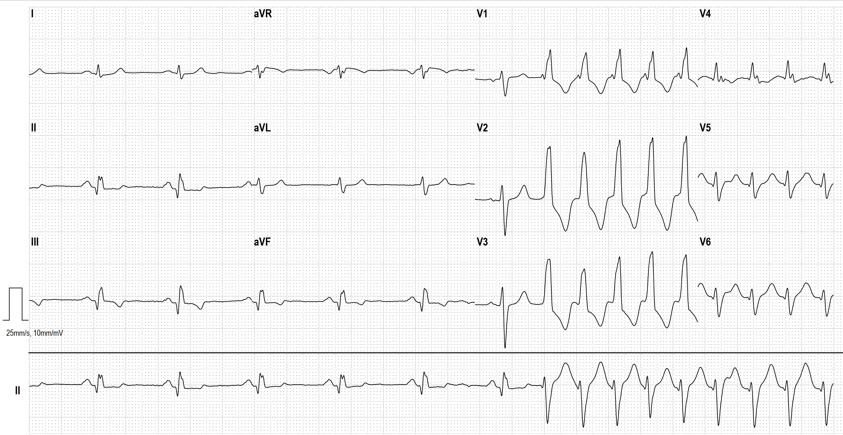
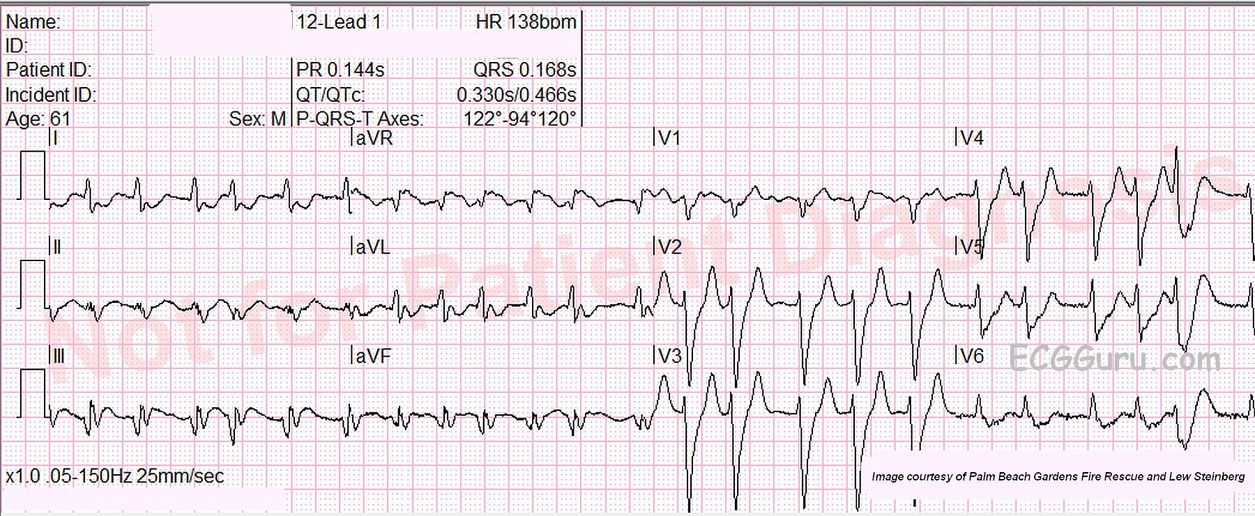
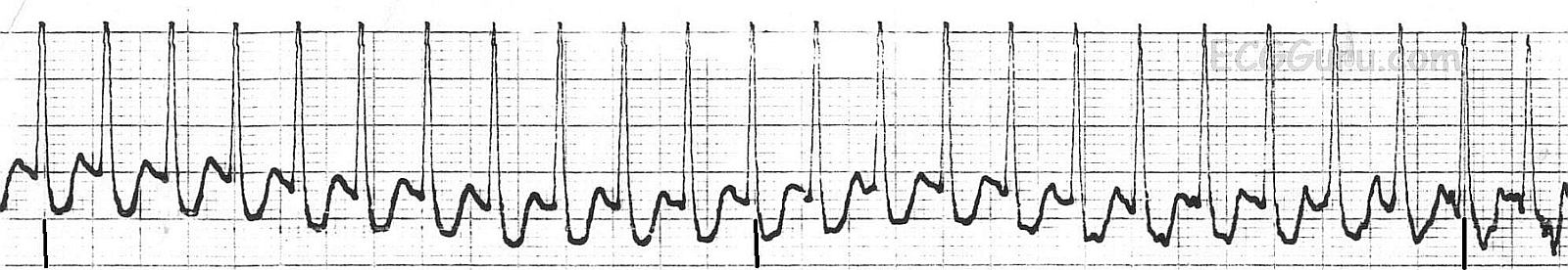
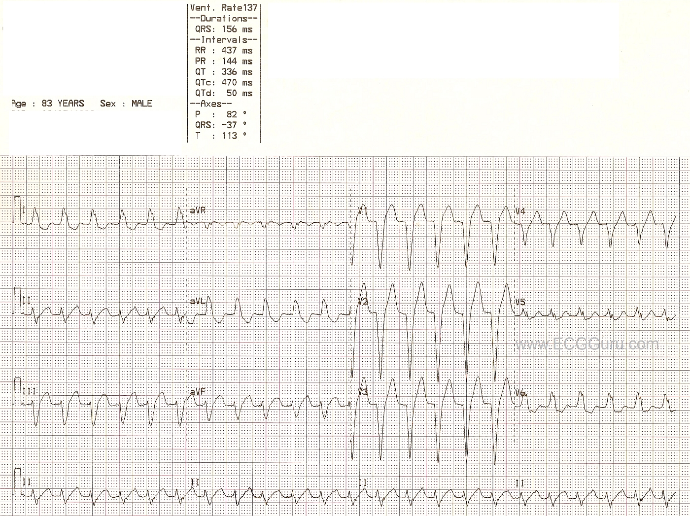
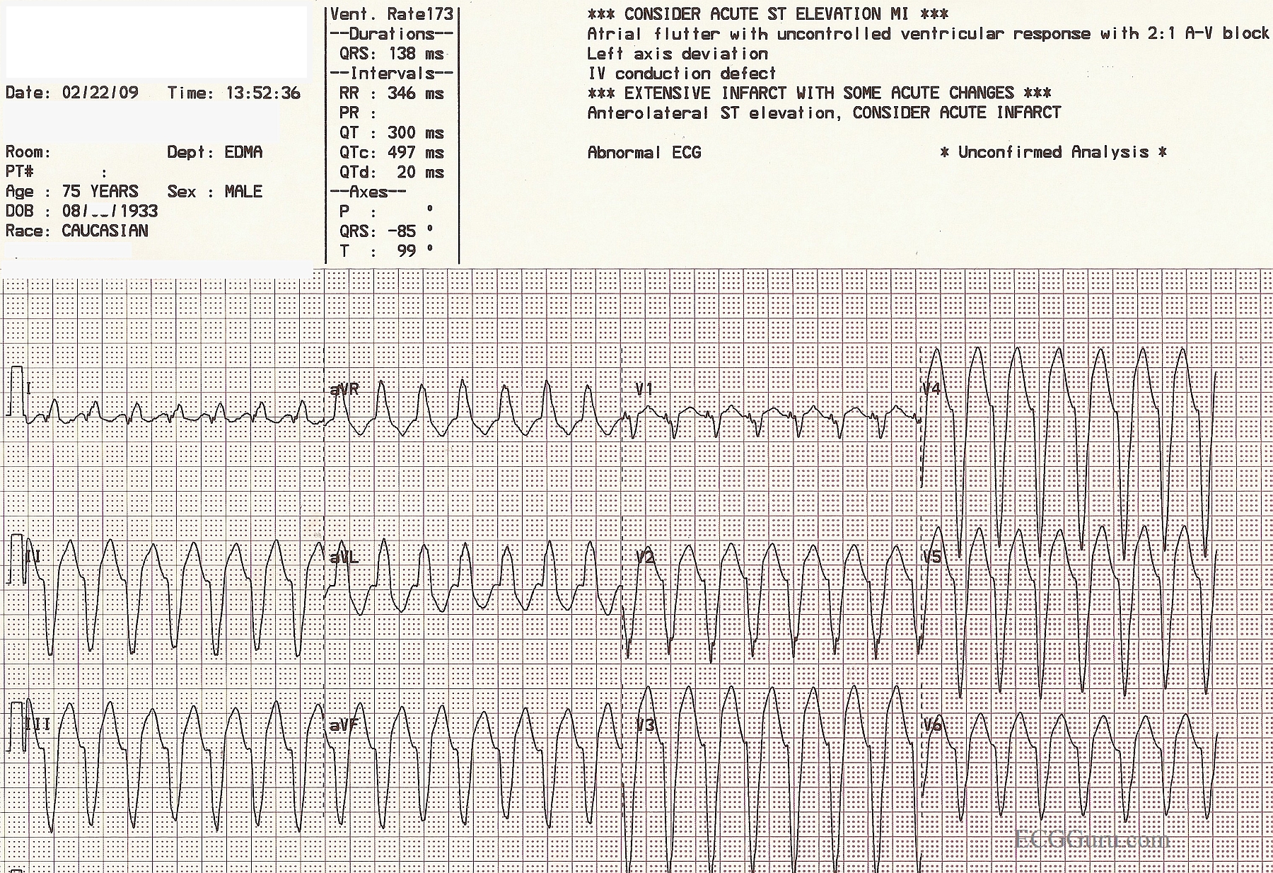
This ECG shows a wide-complex tachycardia with a rate of 137/minute.  No patient information is available other than what is on the ECG.  Here, we will comment for the BASIC LEVEL learner, and allow the ECG Gurus out there to add INTERMEDIATE and ADVANCED level comments.

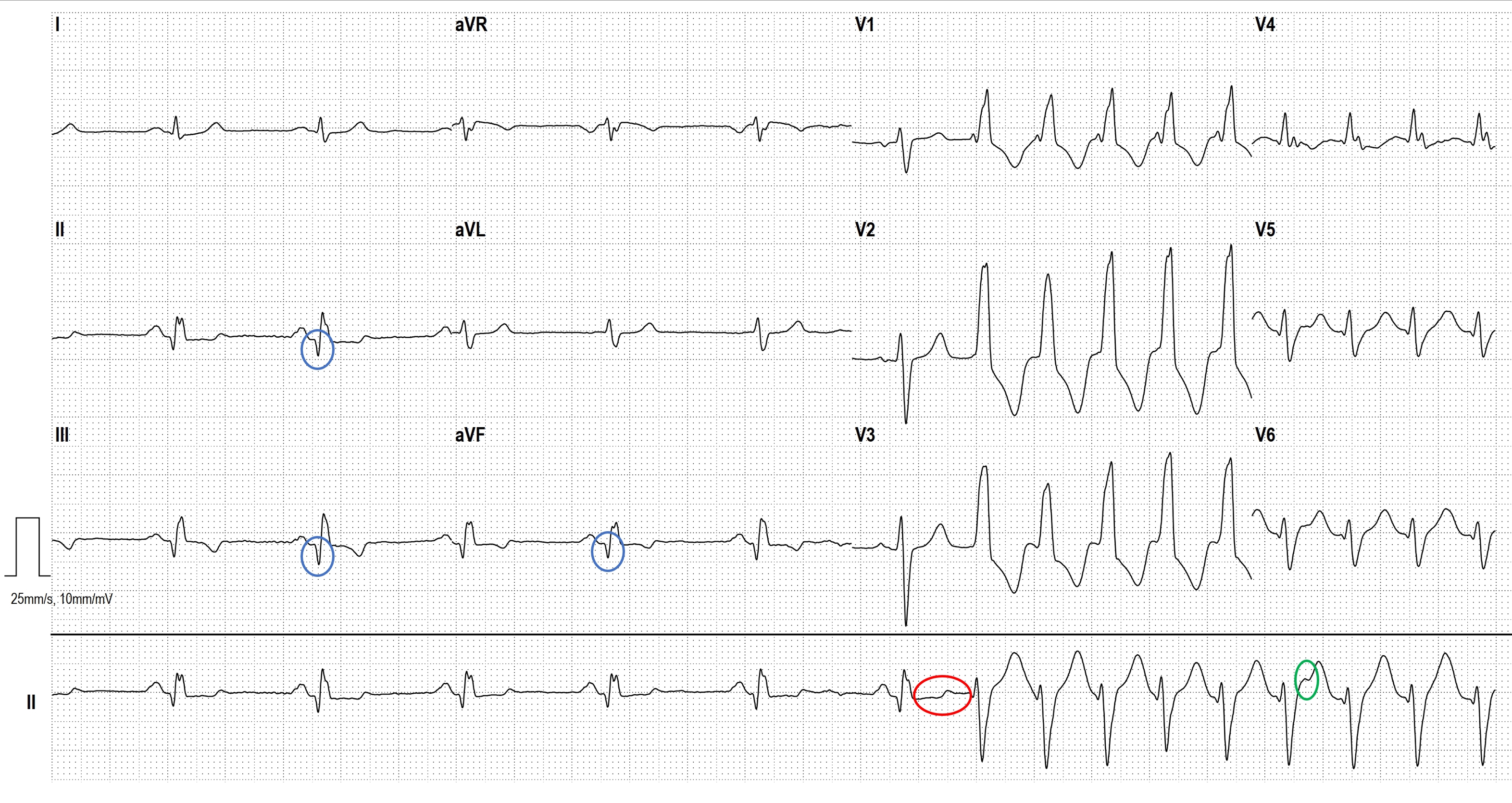
Beginners and emergency workers should ALWAYS be cautioned to treat all wide-complex tachycardias as VENTRICULAR TACHYCARDIA until proven to be something else.  V Tach is a life-threatening dysrhythmia, and there should be no delay in treatment.  That being said, not all WCTs are V Tach.  The most common "mimic" of V Tach is left bundle branch block.  LBBB can appear along with any supraventricular rhythm, including sinus rhythms, atrial tachycardias, atrial fibrillation, and junctional rhythms.  The diagnostic criteria for LBBB are:  wide QRS (.12 seconds or more), supraventricular rhythm, negative QRS in V1 and positive QRS in V6 and Lead I.  This ECG meets the criteria for LBBB, except that the supraventricular rhythm is hard to prove.  On one hand, P waves are not seen - or at least not easily.  On the other hand, the ECG machine has recorded a PR interval and a P wave axis.  What do you think?

Some of the criteria that would favor the diagnosis of V Tach are not present here: precordial concordance (all QRS complexes in V1 through V6 pointing in same direction), V6 with a negative QRS, AV dissociation.  An extreme axis, especially extreme right, would favor V Tach.  This ECG has an abnormal left axis, which is possible in V Tach AND in LBBB.   For a more thorough discussion by ECG Guru Jason Roediger of the criteria for differentiating V Tach from LBBB and other aberrancy, go to this LINK.

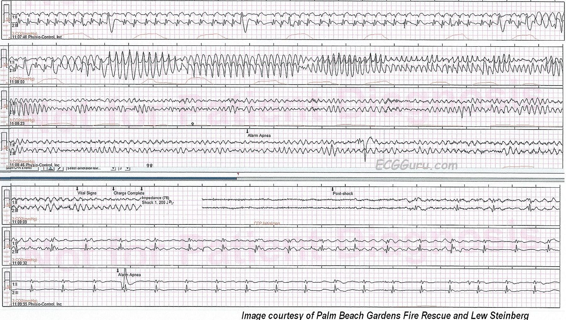
In an emergency setting, WTCs are most often V Tach.  However, should the rhythm turn out to be SVT with aberrant conduction, the usual V Tach treatment protocols would do no harm.  For instance, if the patient is deemed to be "unstable", electric cardioversion is recommended for both V Tach and SVT.  If the patient is stable, the emergency drugs usually recommended are generally safe for both rhythms.

Care should be taken to differentiate SINUS tachycardia from SVT or V Tach.  Sinus tachycardia usually has an obvious physiologic cause (hypoxia, hypovolemia, fear, pain, fever, etc.).  Sinus tachycardia also will be variable in rate, slowing as the cause is alleviated, or speeding if the problem causing the tachycardia becomes worse.  It may require  a long period of observation to determine that the rate is gradually slowing or speeding up.  One would hope to find P waves at some point in sinus tachycardia, and a 12-lead ECG is a great help.

WE LOOK FORWARD TO COMMENTS FROM OUR MEMBERS.

Dawn's picture

Ventricular Tachycardia

This is a good example of ventricular tachycardia with PRECORDIAL CONCORDANCE.  The QRS complexes in the chest, or precordial, leads all point downward.  When the precordial leads are all  negative or all positive in a wide-complex tachycardia, there is virtually a 100% chance that the WCT is ventricular tachycardia.  This ECG shows many characteristics of VT, including the extreme "backwards" axis:  aVR is positive and II, III, and aVF are negative.  Lead I is almost equiphasic.  Also, the lack of a clear BBB pattern and a negative V6 are strongly suggestive of VT.  REMEMBER:  In the treatment of wide-complex tachycardia, the rhythm should be considered VT unless proven otherwise.  This is especially true in unstable patients, patients over 50 years old, and patients with known heart disease.

INSTRUCTORS' NOTE:  We purposely left the machine interpretation on this week's ECG of the Week.  How many errors did the machine make?  This might be a good teaching point for students of all levels.


Dawn's picture

Ventricular Tachycardia

A 66 year old man is complaining of palpitations and chest pain which radiated to his left arm and neck, which lasted 20 minutes, then went away.  Paramedics found him in V Tach.  His BP was 120/80 and his pulse 120/min. He converted to a narrow-complex rhythm while being given amiodarone, but became nauseated.  He returned to V Tach, and his symptoms disappeared.  This patient had an implanted defibrillator, which never went off.  How do we know this is V Tach?

First, ALWAYS consider any wide-complex tachycardia to be VT unless you have proof that it is not.  When symptoms include chest pain, it can be especially dangerous to miss the diagnosis of VT.  Remember, some VT can be asymtomatic, even for prolonged periods of time.

ECG signs that this is VT include: QRS is extremely wide (>.14 sec), no P waves associated with the QRS complexes, negative complexes in V4, V5 and V6.  In fact, this patient has negative "precordial concordance" - all the chest leads are negative. This is a strong sign of VT.  The wide little r wave in V1, greater than .04 sec (one small block) is a strong indicator of VT, as is the delayed nadir of the S waves in V1 through V3 (the slope of the S wave is not steep, indicating a long time to depolarize the ventricles).

For more about V Tach criteria, browse this site and visit the ECG blogs on the Favorites page.  It is a favorite topic among ECG experts.


Dawn's picture

AV Sequential Pacing to Ventricular Tachycardia

This is an interesting ECG for showing students AV sequential pacing and also ventricular tachycardia.  The unusual thing about this ECG is that the V Tach starts at the time the machine begins recording the precordial leads.  This particular ECG machine shows a slight "gap" at the lead change, so we don't see the actual start of the V Tach.  Both rhythms have wide QRS complexes. The pacemaker is pacing the right ventricle, so you will see a wide QRS with a leftward axis, as the impulse spreads up and leftward toward the left ventricle.   The V Tach portion is, of course, limited to the precordial leads, so we cannot plot the frontal plane axis.  But, it meets many of the accepted criteria for ventricular tachycardia, including:  very wide QRS, negative QRS in Lead V6, absence of RBBB or LBBB pattern.   For more on recognizing V Tach in a WCT, go to Ask the Expert at this LINK.    

This is also a very good example of how the interpretation by the machine can be wrong.  Always read the ECG yourself!

Pages

All our content is FREE & COPYRIGHT FREE for non-commercial use

Please be courteous and leave any watermark or author attribution on content you reproduce.