Displaying 1 - 9 of 9
Dawn's picture

ECG Basics: Paroxysmal Supraventricular Tachycardia Converting to Sinus With PACs

This strip shows a supraventricular tachycardia, rate 196 bpm, after adenosine was administered to the patient.  The PSVT breaks, and an irregular rhythm composed of sinus beats and premature atrial contractions ensues.  This is common after medical cardioversion. The patient later settled into a normal sinus rhythm.  The abrupt change from a fast, regular rhythm to a slower, irregular rhythm is evidence that the tachycardia was due to a reentrant circuit, and not sinus tachycardia.

Dawn's picture

Narrow-complex Tachycardia In An Infant

The patient:  This ECG was obtained from a two-month-old girl who was a patient in the Emergency Department.  She had a fever due to a respiratory infection and was dehydrated. She was alert, active, and irritable.

The ECG:  There is a narrow-complex tachycardia at a rate of 194 bpm.  This is faster than the normal range for a two-month-old, which is about 80-160 bpm.  The intervals are all within normal range.  The frontal plane axis, at 145 degrees, is rightward, which is normal for this age. There are prominent, narrow Q waves in the inferior wall leads (II, III, and aVF) and in the left lateral leads (V4, V5, and V6).  There are no Q waves in the high lateral leads (I and aVL).  This is a normal pattern for this age group.   www.sciencedirect.com/science/article/pii/B9781416037743100280

The evaluation of this ECG must be preceded by a thorough evaluation of the patient.  SINUS TACHYCARDIA would be expected in the setting of fever, dehydration, hypoxia, pain or other discomfort. Should the rate fail to gradually return to a normal range after treatment, we would have to consider a reentrant supraventricular tachycardia. Reentrant tachycardias have a SUDDEN ONSET and SUDDEN TERMINATION.

Unfortunately, we do not have follow up on the patient.

Dawn's picture

ECG Basics: Sinus Tachycardia vs. PSVT

Narrow-complex tachycardias can be very confusing to students of basic-level ECG.  There are very many rhythms that fall into the broad category of narrow-complex tachycardia.  We usually further divide them into sinus tachycardia and other "supraventricular tachycardias".  The basic student will want to make this distinction, as well as be able to differentiate atrial fib and atrial flutter from the other SVTs.  The more advanced student will want to go into more detail about which mechanism for supraventricular tachycardia is present.

Just the basics, please.   When the tachycardia is regular, it is most important to determine whether it is a SINUS TACHYCARDIA or a SUPRAVENTRICULAR TACHYCARDIA.  (Yes, we are aware that sinus rhythms are supraventricular, but the term "supraventricular tachycardia" or "SVT" is usually reserved for the fast, regular rhythms that are not sinus.)  So, what clues will be most helpful to our beginner students?

Rate    SVTs tend to be faster than sinus tachycardia.  More importantly, they are fast regardless of the patient's situation.  Sinus tachycardia almost always is reacting to the patient's situation.  For instance, a 22-year-old woman resting in a chair with a heart rate of 150 is likely to have an SVT.  A 22-year-old woman who is running in a 10 k marathon race and has a heart rate of 160 is responding appropriately to an increased need for oxygen and nutrients to her cells. Sinus tachycardia will ususally be 160 or less, and have an obvious reason for being, such as fever, pain, anxiety, exercise, hypovolemia, hypoxia, or drugs.  Unfortunately, many beginning students are told that any narrow-complex tachycardia with a rate of 150 or less is sinus, and over 150 is SVT. While they may be right most of the time, or on the written test they are about to take, this rule should not be applied in "real life".  Sinus rhythms can go over 150, and SVTs can be slower than 150.  So, what other clues should we be teaching beginners?

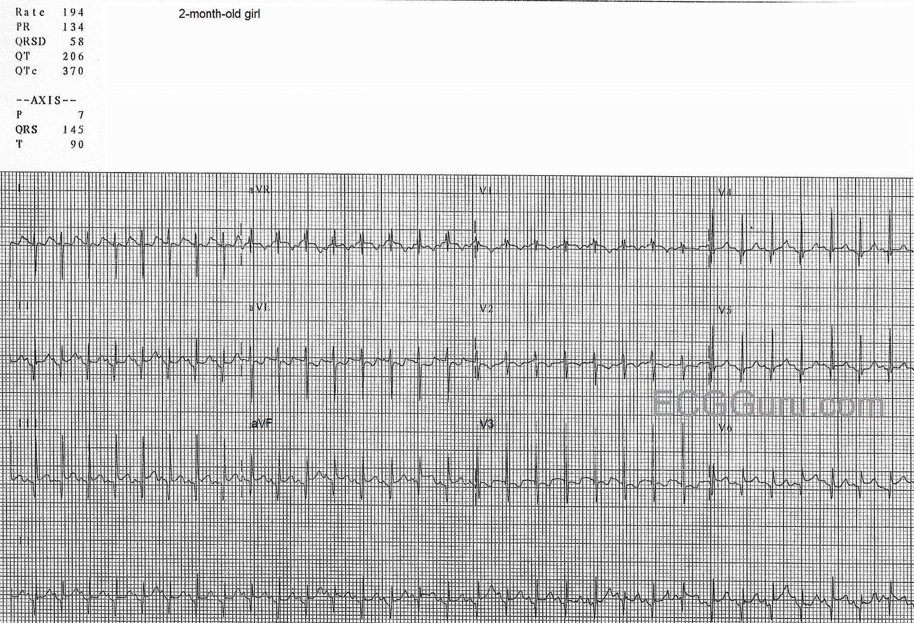
Consider the clinical situation    Look for an obvious cause for sinus tachycardia.  If none is found, strongly consider SVT.  Remember that pediatric patients have faster heart rates, especially infants.  If the strip is on a test, with no clinical information, consider these:

Dawn's picture

Paroxysmal Supraventricular Tachycardia

This ECG is from a man in his 60's who is experiencing chest discomfort and palpitations.  The onset of the rapid heart rate and the symptoms was sudden, while he was at rest.  The rate did not slow when he was placed on oxygen, given IV fluids, and rested further. The rate is 177 / min.  

The rhythm is AV nodal reentry tachycardia (AVNRT), which is one of the rhythms that falls into the category of paroxysmal supraventricular tachycardia (PSVT).We can see signs of retrograde P waves in some leads (II, III, aVF, V1).  AVNRT is caused by a reentry circuit in the AV node.


Some instructors teach students that sinus tach is approximately 100-150 per minute, and atrial tach is usually 150-250 per minute.  If students only learn about differentiating these two rhythms by the rate difference, it will cause later problems.  Of course, there is actually an overlap in rates between the two rhythms.  For example, a febrile, dehydrated infant could easily reach this rate and be in sinus rhythm.  A young, healthy person on a treadmill could, too.  Clues to the ectopic origin of this rhythm are:  sudden onset (unfortunately, not witnessed here), regular rhythm with unwaivering rate, and the patient's situation (symptoms while at rest, no obvious reason for sinus tach).  Of course, we need to teach to the level of our students' abilities.  Consider whether they just memorizing rhythms criteria now, or are they learning about re-entry?

Dawn's picture

ECG Basics: Paroxysmal Supraventricular Tachycardia Treated With Adenosine

This series of ECG rhythm strips shows a paroxysmal supraventricular tachycardia successfully treated with adenosine.  The patient was complaining of a rapid heart rate and palpitations, but was hemodynamically stable.  It is not known whether any parasympathetic stimulation, such as a Valsalva maneuver or carotid sinus massage, was used initially.   

The first rhythm strip shows a PSVT, presumably AV nodal reentrant tachycardia, at a rate of about 215 per minute.  (We originally indicated a rate of 240 per minute, but this was a typo). Using the simplist method of determining rate, the six-second method, we see 21, but almost 22, QRS complexes in six seconds. Differential diagnosis would include sinus tachycardia, but this rate is too fast for sinus tach, especially in a resting patient.  Also, sinus tach would slow down as the patient is rested or made more comfortable, and this rate did not vary.  Also, when confronted with a supraventricular tachycardia, one should also consider atrial flutter and atrial fibrillation.  This is somewhat slow for atrial flutter with 1:1 conduction, and that rhythm is much more rare than AVNRT. It is too regular for atrial fibrillation.  So, we are left with the probable diagnosis of paroxysmal supraventricular tachycardia.  The “paroxysmal” part is presumed since AVNRT has an abrupt onset, and the patient’s symptoms started suddenly. 

The second rhythm strip shows what happened after adenosine was administered.  The patient received first a 6 mg dose, rapid IV push.  When that was not effective, he received 12 mg rapid IV push.  The rhythm strip is typical of the first minute or so after adenosine administration.  Adenosine can cause transient AV blocks, escape rhythms, and ectopic irritability.  The half-life of adenosine is only 6 seconds, so the dysrhythmias and uncomfortable symptoms are short-lived.  In this strip, we see frequent PVCs and runs of V tach.  

Dawn's picture

Supraventricular Tachycardia

This ECG shows AV nodal reentrant tachycardia in an elderly man.  Clinical information is not available.  AVNRT is the rhythm most often associated with the term, "supraventricular tachycardia".

Although we can't see the beginning of this rhythm, one of the identifying features of SVT is an abrupt (paroxysmal) onset.  In patients with AVNRT, there are two pathways in the AV node, a pathway with fast conduction and a long refractory period, and a pathway with slow conduction and a short refractory period.  Normal sinus impulses travel down the fast pathway and into the ventricles, but also start up the slow pathway in a retrograde direction. The retrograde impulse and the normal impulse traveling down the slow pathway collide, cancelling each other out.  If a PAC occurs, it will travel down the slow pathway while the fast pathway is still refractory.  By the time the impulse reaches the end of the slow pathway, it finds the fast pathway no longer refractory, and travels back up to the atria.  This forms a circular movement (circus movement) of the impulse, and it repeats itself rapidly until interrupted.  When each impulse reaches the ventricles, it travels into the interventricular conduction system and causes ventricular depolarization and contraction, usually at a rate of 140 - 250+.  Unlike sinus tachycardia, AVNRT does not adjust its rate according to the needs or activity of the patient.

There are many forms of supraventricular tachycardia, and they are not always easy to differentiate based on ECG criteria alone.  AVNRT of the type described above is the most common PSVT in structurally normal hearts.  For more information on supraventricular tachycardia, go to Life in the Fast Lane.  For a discussion on clinical management, we recommend Dr. Grauer's ECG Video 6 - Rhythm Mgmt-Part 3.

Dawn's picture

ECG Basics: Paroxysmal Supraventricular Tachycardia

This two-lead rhythm strip clearly shows the transition from normal sinus rhythm to a paroxysmal supraventricular rhythm.  In this case, the arrhythmia is AV nodal reentrant tachycardia, AVNRT.  The rate of the first rhythm, NSR, is around 75 per minute.  The fourth beat on the strip is a PAC which initiates the paroxysm of tachycardia lasting 12 beats.  The arrhythmia terminates spontaneously at that point.  The tachycardia rate is about 150/min.

The topic of supraventricular tachycardias can be a very complex one to teach.  For an excellent example of a concise lesson geared toward Primary Practice physicians, go to Dr. Grauer's VIDEO - Part III of his Arrhythmia series.

To cover the important points for the beginner-level student:

  *  It can be difficult to determine a rhythm is SVT if the rhythm is near 150 bpm and you DON'T see the beginning or end of the arrhythmia.  If the onset (or offset) is sudden, then this is not a sinus rhythm.  The sinus node speeds and slows more gradually - it doesn't change rates in one heartbeat.  This strip has an excellent view of BOTH the onset and the offset.

  *  The faster the rate, the more likely we are looking at a PSVT rather than sinus rhythm.  If a sinus tachycardia exists, we can almost ALWAYS see the reason for it in the patient's clinical situation.  We may see fever, dehydration, bleeding, fear, pain, exercise.  Therefore, a patient at rest with a rate of 150 would be suspect for PSVT.  A patient on a treadmill for 5 minutes would be considered to have a sinus rhythm.

  *  Any patient with a rate around 150 per minute should be evaluated for ATRIAL FLUTTER with 2:1 conduction.  Atrial flutter often conducts at that ratio, because a rate of 150 is fairly easy for the AV node to conduct, whereas the instrinsic rate of atrial flutter (250-350) is not.  A 12-lead ECG makes it easier to search for tell-tale flutter waves.

Dawn's picture

ECG Basics: Paroxysmal Supraventricular Tachycardia Converted With Adenosine

Today, you get THREE strips for your basic classes!  The first shows a PSVT - paroxysmal supraventricular tachycardia at a rate of about 220/minute.  The QRS complexes are narrow, and the rhythm is regular.  In the second strip, we see the moment of conversion after a dose of 6 mg. of adenosine was administered by rapid I.V. infusion.  The re-entry cycle is broken, and the patient experiences many PVCs, including groups of two, three, and even four in a row.  Soon, however, the rhythm settles into sinus rhythm with PACs, and later, just sinus rhythm (not shown).  For your more advanced students, the second strip shows the PVCs interacting with the underlying sinus rhythm.  There are several fusion beats with varying degrees of fusion, and one can sometimes see a sinus P wave just before a PVC.  None of this is clinically significant in this patient, because the ventricular ectopy was a side effect of the treatment, and was self-limited.  It is a great strip for teaching, though! 

Dawn's picture

ECG Basics: Supraventricular Tachycardia

This strip is from a patient who experienced a sudden onset of palpitations and rapid pulse while at rest.  It shows a narrow-complex tachycardia, specifically a paroxysmal supraventricular tachycardia.  The subject of supraventricular tachycardias is a fascinating one, and is covered extensively throughout this website.  The mechanisms of SVT are many, and can be complex for the beginning student to understand.  Search the search terms on the left side of the page for entries from ASK the EXPERT and JASON's BLOG for more advanced information about SVT.

For the beginner, it is important to teach the difference between sinus tachycardia and "supraventricular tachycardia".  Of course, sinus tachycardia IS supraventricular - but current convention has us using the term "SVT" for atrial or junctional tachycardias, and especially for reentrant tachycardias.  Beginner students should understand the function of the sinus node, and it's ability to control the heart rate, based on direction given by the nervous system.  The sinus node increases and decreases the rate incrementally, or more gradually than the onset and offset of a reentrant tachycardia.  The appearance of a sudden onset of regular tachycardia following a PAC, producing a rhythm with a distinctly faster rate than the original sinus rhythm, is a sure sign of SVT.  When the onset or offset are caught on the rhythm strip, our job is SO much easier!

The heart rate helps with the diagnosis.  SVTs tend to be faster than sinus tachycardias.  SVTs tend to be faster than 150/min, while sinus rhythms TEND to be slower than 150.  And patients with sinus tachycardia usually have a readily determined reason for the tachycardia, such as fever, pain, fear, hypovolemia, hypoxia, or exertion.  So, a patient on a treadmill for an exercise stress test might very well have a heart rate over 150 / min.

When your students master the understanding of the different behaviors of the sinus node and the reentrant rhythms, and how important patient presentation is to the diagnosis, you will want to add atrial flutter and atrial fibrillation.  When these are mastered, be sure to remind your students that atrial flutter can conduct 2:1, and will mimic sinus tach.  A good rule of thumb is: Under 150/min.:  look for sinus tach first.  Around 150 / min.: look for atrial flutter with 2:1 conduction.  Over 150/min.: suspect PSVT.   


All our content is FREE & COPYRIGHT FREE for non-commercial use

Please be courteous and leave any watermark or author attribution on content you reproduce.