Displaying 1 - 8 of 8
Dawn's picture

Complete Heart Block or High Grade AVB?

The patient:  This ECG was obtained from a 91-year-old woman who was complaining of weakness.  Unfortunately, we have no other information. 

The ECG:  This ECG has something for your basic students, and even more for the more advanced learners.  The first thing  that anyone should notice is the slow rate.  The ventricular rate is around 35 bpm, and regular.  If the patient is showing signs of poor perfusion, we would stop here and prepare to increase the rate with a temporary pacemaker (transvenous or transcutaneous). Why is the rate so slow?  There is no P wave in front of each QRS, so this is not sinus bradycardia.  Rather, we see P waves at a rate of approximately 100 bpm, wit a very regular rhythm.  Beginners should “march out” the P waves with calipers or by marking a straight edge piece of paper.  There are 15 P waves on this ECG – some are buried within QRS complexes (QRS #3) or T waves (QRS #4).

 Because there are two distinct, regular rhythms, but they do not track with one another, we know this is possibly third-degree AV block (complete heart block).  Another clue is that there are no steady, repetitive PR intervals, which means there is no relationship between the atrial rhythm and the ventricular rhythm.

 For more advanced learners, it is helpful to try to identify the origin of the escape rhythm.  If it is junctional, the AV block is above the junction.  If the escape is ventricular, the AV block is below the junction.  A junctional rhythm is usually between 40 – 60 bpm, with a narrow QRS.  Ventricular escape rhythms are usually less than 40 bpm and with wide QRS complexes.   This ECG will be a little challenging on this front, because the rhythm has some characteristics of junctional rhythm and of ventricular rhythm.

Dawn's picture

Shark Fin Pattern

The Patient:  This ECG is from a 59-year-old woman who was found by the EMS crew to be unresponsive, with agonal respirations at about 6 breaths per minute. Her pulse was thready at the carotid, and absent peripherally. Her skin was pale, cool, and mottled.  Her BP via the monitor is 81/40, peripheral pulses not being palpable.  An initial rhythm strip showed sinus rhythm at 75 bpm with right bundle branch block and ST elevation.

The patient’s husband gave a history of “difficulty breathing” since sometime this morning, alcohol dependence, hypertension, tobacco use, and insomnia. He said she had been drinking heavily for several weeks.

She was immediately ventilated and intubated, and an intraosseous infusion line established.  A12-lead ECG was done, and it showed a dramatic change in the rhythm and ST segments over the initial strip. She was transported to a nearby hospital with CPR support. She achieved return of spontaneous circulation (ROSC) at the Emergency Department, after having three doses of epinephrine.  Follow up with the ED physician revealed that the patient had suffered a massive gastrointestinal bleed.  This patient, due to loss of a critical amount of blood, had low blood pressure and very poor perfusion, which resulted in damage to her heart (and possibly other organs as well). I do not have further follow up, but will update this if I receive more information. 

Dawn's picture

Inferior Wall M.I. With Wide QRS and Complete AV Block

This ECG is from a 66-year-old woman who called 911 for a complaint of chest pain for the past four hours. She also complained of nausea, vomiting, and diarrhea for that time. She was pale and diaphoretic, and her BP was 77/43 sitting up, improving to 90/54 reclining. She denied “cardiac” history.  Her medications included:  aspirin, an SSRI, cilostazol, amlodipine, umeclidinium and vilanterol inhaler, atorvastatin, levothyroid, and metoprolol. We don’t have a previous ECG.  The EMS crew followed their chest pain protocol and delivered the patient to a facility with an interventional cath lab, but they did not designate a “STEMI Alert” because of the wide QRS.  It is their protocol to use the term “STEMI Alert” only when no M.I. mimics, such as left bundle branch block, are present. 

What does this ECG show?     There is an underlying sinus rhythm at 75 bpm.  There is AV dissociation, with regular, wide QRS complexes at a rate of 44 bpm.   This meets the criteria for complete heart block (third-degree AV block).  The morphology of the QRS complexes meets the criteria for left bundle branch block (wide, upright in Leads I and V6, negative in V1).  At a rate of 44 bpm, several options for this escape rhythm are possible:  1)  junctional escape with LBBB, 2) junctional escape with intraventricular conduction delay due to AMI,  and 3) idioventricular escape rhythm.   Also, in the presence of IWMI, AV node ischemia is very likely, resulting in AV blocks at the level of the AV node.  CHB at the AV node would result in junctional escape rhythm, and CHB below that, in the fascicles of the bundle branches, would result in idioventricular escape. The issue for this patient, and ANY patient, is cardiac output, and we see several reasons for cardiac output to be lower:

·         Wide QRS

Dawn's picture

Paced Rhythm Following AV Node Ablation

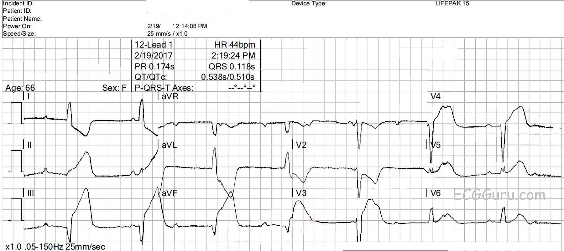
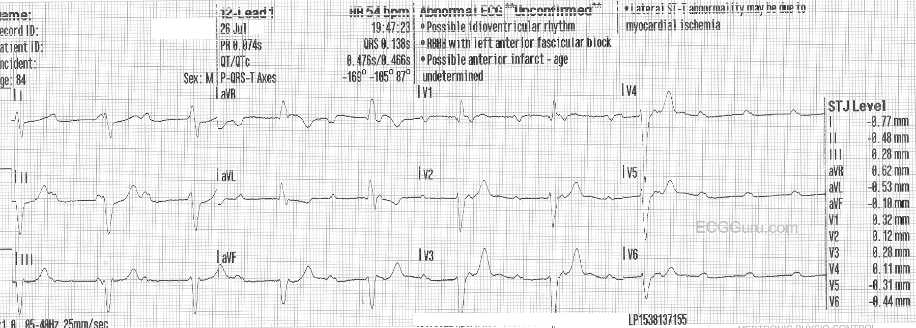
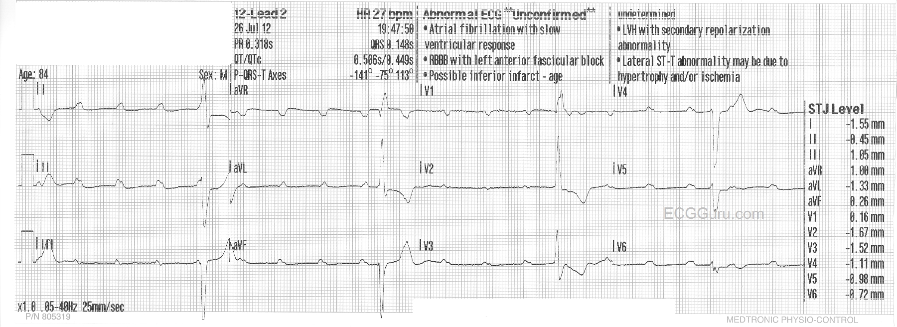
This ECG is taken from a woman who had suffered for several years with intractable intermittent atrial fibrillation. She had tolerated medications poorly, and several attempts at electric cardioversion had resulted in only temporary relief. Ultimately, she chose to undergo AV node ablation.  In the electrophysiology lab, her AV node was destroyed, preventing the atrial fib impulses from penetrating into the ventricles.  This resulted in a “man-made” complete AV block.  A pacemaker was implanted in the EP lab.  When she is in atrial fibrillation, the fibrillatory waves of the atria INHIBIT the atrial pacing electrode from firing, so she has no paced P waves at that time.  The right ventricular pacing electrode functions without inhibition, and makes a wide QRS complex with a leftward axis deviation (normal for RV pacing). 

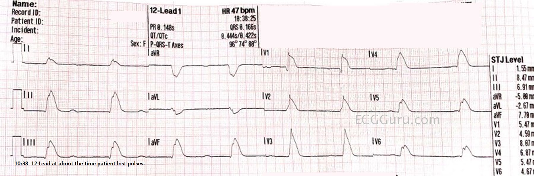
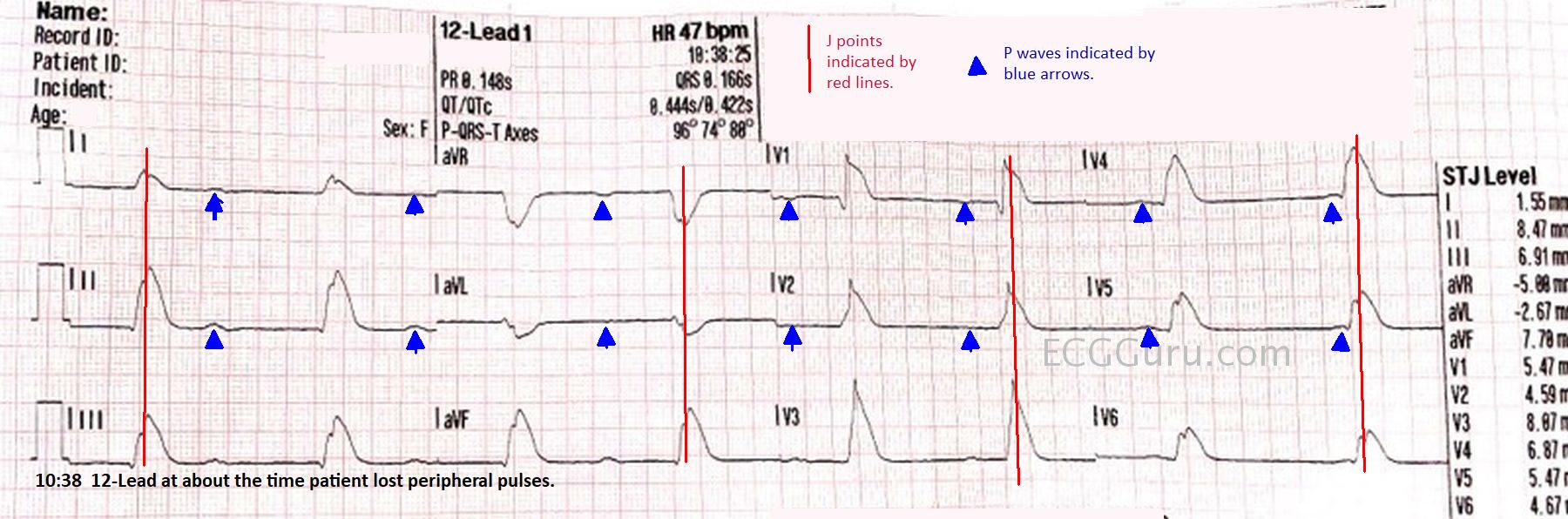
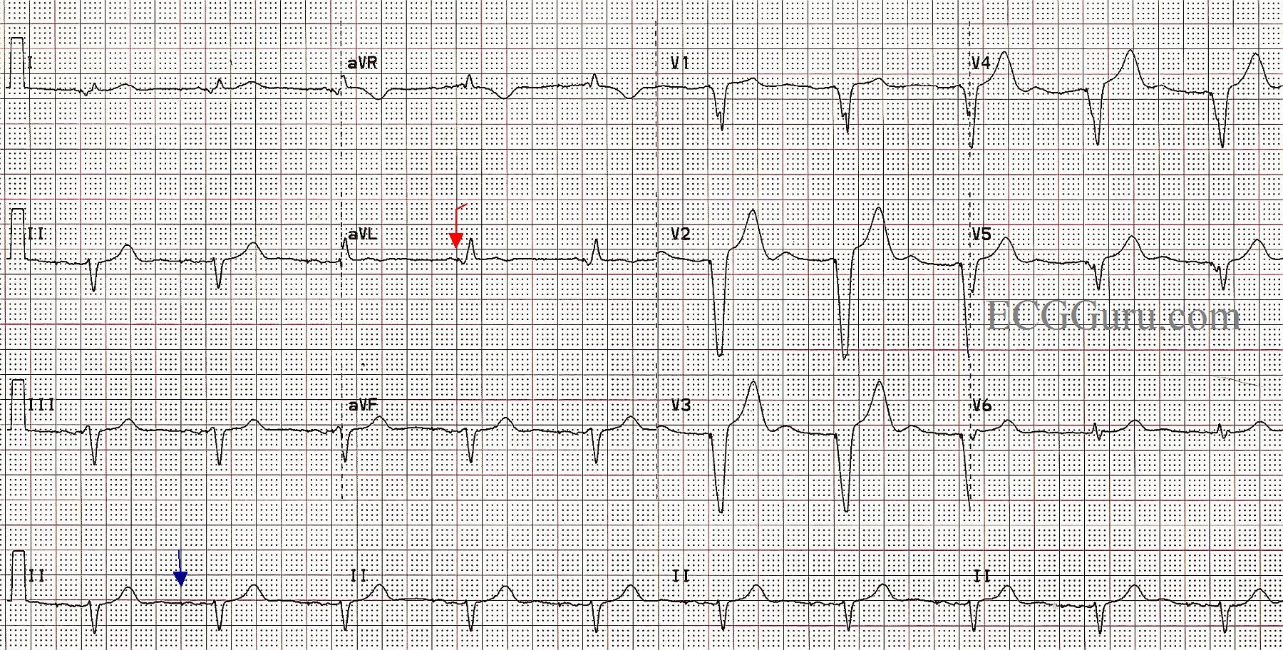
In this ECG, we see the patient WITHOUT atrial fib, and the pacemaker is pacing the atria AND the ventricles, in a sequential fashion.  The spikes are very hard to see, as this is a “bipolar” pacemaker, which makes much smaller spikes than a “unipolar” pacemaker.  Some ECG machines will automatically enhance the spikes, but this one did not.  We have marked a “sample” atrial spike in blue for you and one of the ventricular spikes in red.  Each beat on this ECG actually has appropriately-timed atrial and ventricular stimuli (spikes), and the patient has optimized cardiac output provided by the “atrial kick”. A P wave occurring just before a QRS indicates that the ventricles are filling from the forceful contraction of the atria.  This provides much better filling than when the atria are not beating or are fibrillating. 

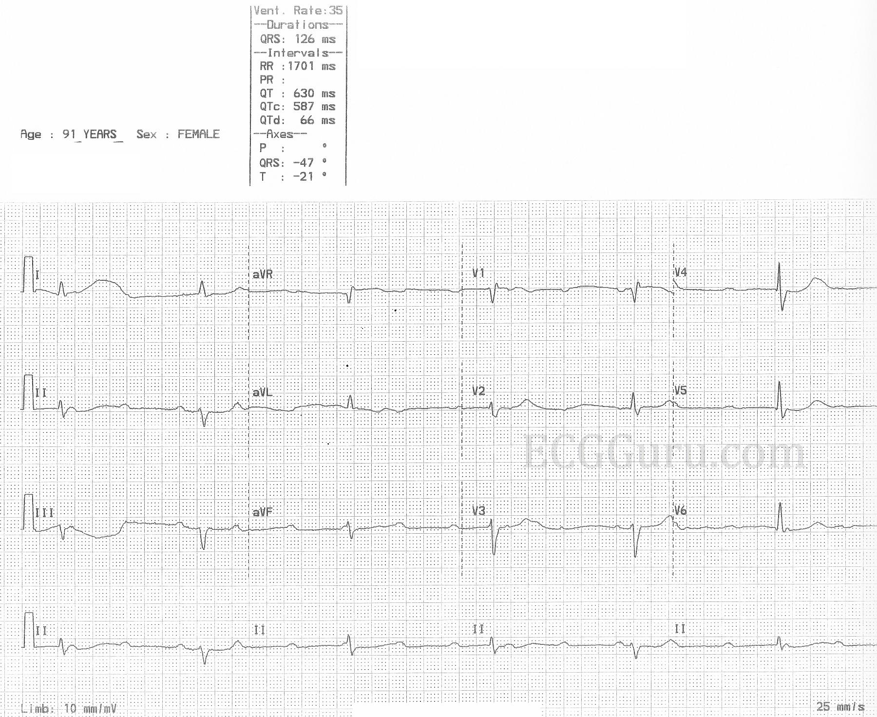
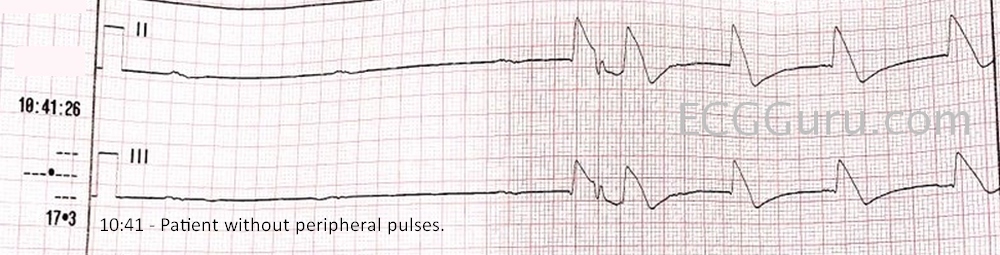
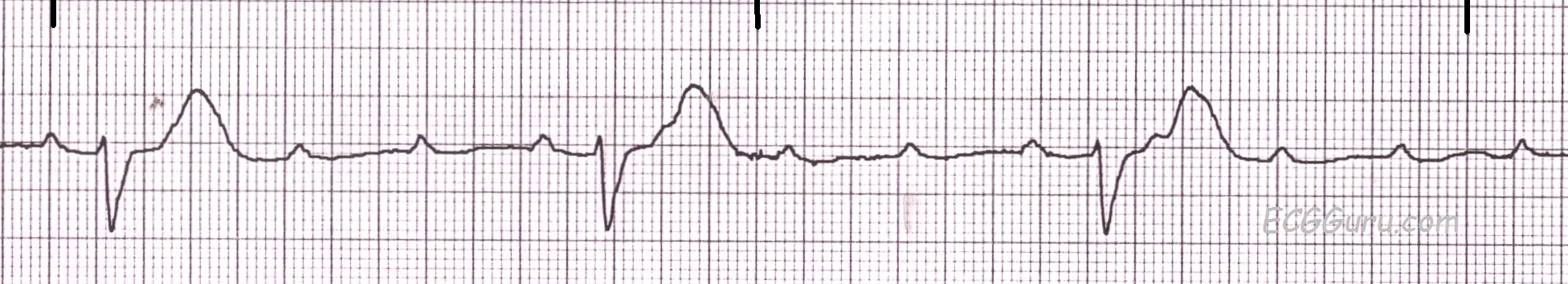
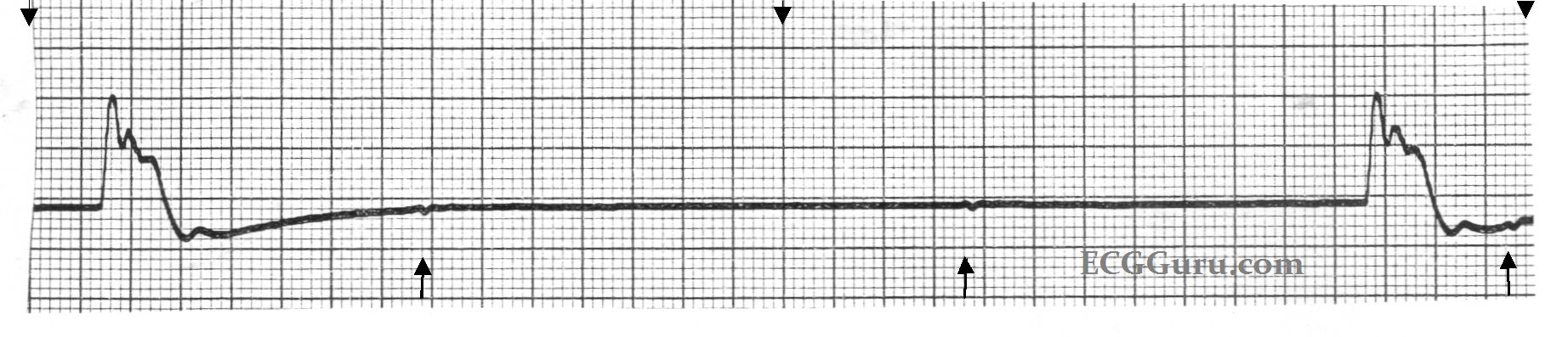
This is a good ECG to use to show your students how we can recognize a paced rhythm without being sure of the spikes.  Of course, without other evidence (patient history and exam), we can’t know for sure that this is a paced rhythm, but the steady, normal rate, wide complexes, and left axis deviation are signs of RV pacing.  Look for negatively-deflected QRS complexes in II, III, and aVF and positive QRSs in aVL and aVR. 

When pacing only one ventricle, the impulse travels relatively slowly through the other ventricle, resulting in wide QRS complexes.  This then results in SECONDARY ST-T WAVE CHANGES.  Wherever the QRS is positive, you will normally see some ST depression and T wave inversion.  In leads with negative QRS complexes, the opposite is true, and you will see ST elevation and upright T waves.  This can make evaluation of ST segments for acute M.I. difficult.


Dawn's picture

ECG Basics: Third-degree AV Block, Complete Heart Block

This rhythm strip shows third-degree AV block, also called complete heart block or complete AV block.  The P waves are from the sinus node, and are regular at a rate of about 120/min. (Sinus tachycardia). This is a good strip for showing your students how to "march out" the P waves to find the ones that are hidden behind QRS complexes or T waves. Knowing that the P waves are regular, it is easy to find the hidden ones.

The QRS complexes are wide at 0.14 seconds, and regular, with a rate of about 28/min.  On first glance, it APPEARS that there are PR intervals.  That is, it appears that some of the P waves are conducting. If you measure the PR intervals carefully, you will note that they are NOT equal.  There is no connection between the P waves and the QRS complexes - this strip has just caught them near each other.  If we ran the strip longer, we would see the PR intervals "come apart", proving they are not real.  The QRS complexes are coming from an IDIOVENTRICULAR ESCAPE RHYTHM.  They are regular, wide, have no P waves associated with them, and the rate is below 40 bpm.

Patients with CHB that results in a very slow heart rate sometimes need emergency treatment aimed at increasing the rate.  When the escape rhythm is idioventricular, it is assumed that the AV block is located below the AV node, and emergency temporary pacing is often the method of choice.  In fact, a permanent implanted pacemaker is almost always needed.  When the AV block is located in the AV node, the escape rhythm will be junctional (narrow QRS complexes, rate about 40-60 bpm).     


Dawn's picture

ECG Basics: Idioventricular Escape Rhythm

This six-second monitor strip was from a patient who was designated "Do Not Resuscitate", and whose heart rhythm was slowing dramatically.  It shows an idioventricular escape rhythm, with very wide QRS complexes and only two complexes in six seconds. (The top arrows mark three-second segments.)  If you look closely at the points marked by the lower arrows, you will see small, uniform, regular P waves.  The mechanism leading to this agonal rhythm was complete heart block.  A longer strip would show the P waves as all alike, and fairly regular, but slowing.  

Dawn's picture

High-grade AV Block vs. Complete Heart Block

This week's ECG of the WEEK was donated to us by Sebastian Garay. These two ECGs were obtained less than 30 seconds apart from an 84 year-old man who called fire-rescue because he felt dizzy and fell.  He was not injured in the fall, and his vital signs remained stable, with an adequate BP.  These two ECGs were obtained prior to arrival in the Emergency Dept.

The first one shows a sinus rhythm at about 110/min.  There is a complete heart block (third-degree AV block), and the escape rhythm is a wide-complex rhythm at a rate of about 54/min and slowing severely toward the end.  The second ECG was taken less than 30 seconds after the first, and shows a significantly slower escape rhythm rate at 27/min., while the sinus rate increases to 120/min.  The change is sinus rate is likely an attempt by the nervous system to compensate for the lower cardiac output as the ventricular rate slows. The escape rhythm is not only slower, but there are some changes in the QRS morphology from the first ECG.

For your basic students, this ECG serves to demonstrate the AV dissociation seen in complete heart block.  It is easy to "march out" the P waves, and see that some of them are "hiding" in the QRS comlexes.  It also shows how quickly a rhythm can change rates.

For your more advanced students, you will want to have a discussion about escape rhythms.  This one initially has a fairly fast rate, suggesting junctional origin. The QRS morphology is of the right bundle branch type, with left anterior fascicular block.  However, ventricular rhythms originating from the posterior fascicle region can have the "RBBB / LAFB" morphology.  If this escape rhythm is fascicular (ventricular) in origin, it is an accelerated idioventricular rhythm.  The second escape rhythm appears very similar to the first, with the very noticeable exceptions of QRS morphology, especially in V1 and V2, and the rate.

This patient was given Atropine in the ED, with no change to the rhythm.  We do not know what transpired after that, but suspect a pacemaker was in his future. 

We look forward to comments from our members about these two very interesting ECGs.


Dawn's picture

Third-degree AV Block (Complete Heart Block)

This 84-year-old man called 911 because he felt dizzy and fell.  He was not injured in the fall, but the paramedics noted a slow pulse. He denied significant medical history. The initial ECG showed sinus rhythm at about 80 bpm and AV dissociation with an apparent acellerated idioventricular rhythm at about 40 bpm.  Less than one minute later, he has developed a complete heart block with an idioventricular escape rhythm less than 30 bpm.  The escape rhythm speeds slightly toward the end of the strip.  He retained stable vital signs and adequate perfusion during transport.  It is presumed that he was scheduled for an implanted pacemaker.  It is interesting to note the machine's interpretation, and it reminds us to always interpret the ECG ourselves.   Thanks to ECG Guru member, Sebmedic, for his contribution of this ECG. 

All our content is FREE & COPYRIGHT FREE for non-commercial use

Please be courteous and leave any watermark or author attribution on content you reproduce.