Displaying 1 - 3 of 3
Dawn's picture

ECG Basics: Retrograde P Waves

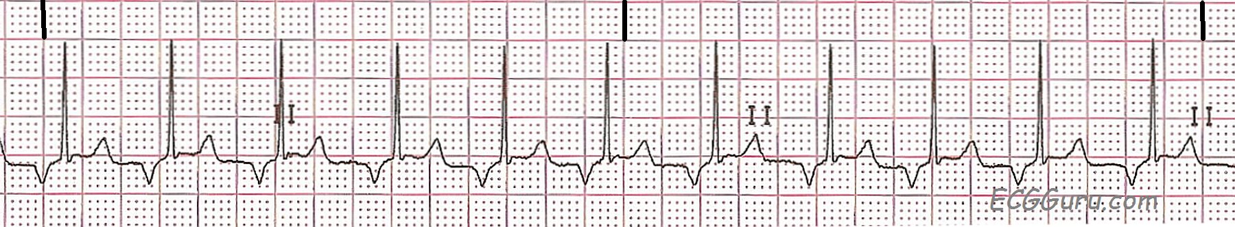
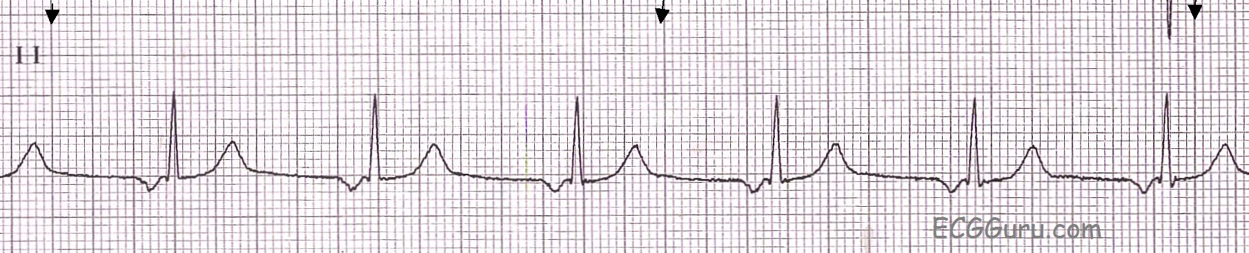
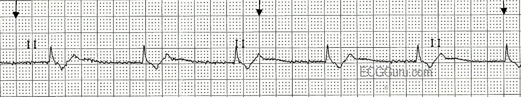
This Lead II rhythm strip shows a regular rhythm with narrow QRS complexes and retrograde P waves.  The strip was taken from a nine-year-old girl.  The rate is about 110 per minute and the PR interval is .12 seconds (120 ms).

When retrograde conduction is seen in the atria, it is often assumed that the rhythm is originating in the junction.  When a junctional pacemaker is initiating the rhythm, the atria and ventricles are depolarized almost simultaneously.  This can produce a P wave in front of the QRS with a short PR interval, during the QRS, or after the QRS.  Sometimes, in junctional rhythm, a block prevents the impulse from entering the atria, producing NO P wave.  Junctional rhythms are usually slow "escape" rhythms, but can be accelerated or tachycardic.

The fact that this rate is 110 / minute and the PR interval is normal at .12 seconds, we should consider that this rhythm could also be from an ectopic pacemaker low in the atria.  From this low starting point, the impulse will travel backward, in a "retrograde" fashion, through the atria, producing a negatively-deflected P wave in Lead II.

We do not have clinical data on this patient, and so do not know what possible causes of arrhythmia might be present, and what the expected rate should be in this situation.  

Dawn's picture

ECG Basics: Junctional Rhythm

A basic rhythm strip showing junctional rhythm in Lead II.  The junctional pacemaker is located between the atria and the ventricles, and the resulting P wave is caused by retrograde conduction through the atria.  This causes the P wave to be negatively deflected in Lead II.  In junctional rhythms, the P wave can occur just before the QRS, during the QRS, or after the QRS, or may not be seen at all.  If the P wave occurs before the QRS, the PR interval is usually short, reflecting the fact that the atria and the ventricles are depolarized almost simultaneously.  In this example, the PRI is .12, on the short side of normal.

The junctional pacemakers have a slow intrinsic rate so that the sinus node can remain in control of the heart's rate under normal circumstances.  If the sinus rate drops below the intrinsic rate of the junctional pacemaker, the junction will take over control of the heart.  An idiojunctional rhythm is generally between 40 and 60 bpm.  In this example, it is about 63 bpm.

Dawn's picture

ECG Basics: Junctional Rhythm

This rhythm strip illustrates a junctional escape rhythm.  The sinus rhythm has slowed or stopped, and the junctional tissue has taken over as the pacemaker of the heart.  The "junction" is loosely defined as the area between the AV node and the Bundle of His.  The intrinsic rate of the pacemaking tissue in this area is 40 - 60 beats per minute.  This slow rate is usually overridden by the sinus node, and the junction is not allowed to express itself as a pacemaker.  Should the sinus node fail or fall below the junctional rate, the junction "escapes" and takes control of the heart.  The QRS complex in junctional rhythm will normally be narrow, because the impulse follows the bundle branches down through the ventricles in a normal fashion, resulting in quick and normal ventricular depolarization.  If the QRS complex is wide in a junctional rhythm, there is another, separate cause, such as bundle branch block.

 If the junctional impulse is able to penetrate the AV node and depolarlize the atria, the P wave will be deflected downward in Leads II, III, and aVF, as the impulse is travelling in a retrograde direction (backward).  The P wave could end up slightly before the QRS, during the QRS, or after the QRS. In this strip, the P waves are seen after the QRS complexes.

All our content is FREE & COPYRIGHT FREE for non-commercial use

Please be courteous and leave any watermark or author attribution on content you reproduce.