
Submitted by Dawn on Sat, 12/28/2013 - 18:59
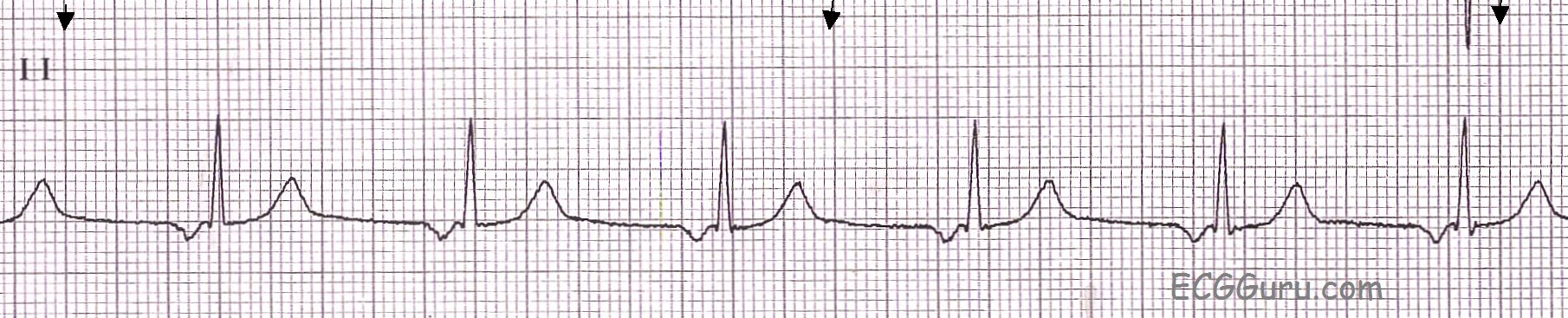
A basic rhythm strip showing junctional rhythm in Lead II. The junctional pacemaker is located between the atria and the ventricles, and the resulting P wave is caused by retrograde conduction through the atria. This causes the P wave to be negatively deflected in Lead II. In junctional rhythms, the P wave can occur just before the QRS, during the QRS, or after the QRS, or may not be seen at all. If the P wave occurs before the QRS, the PR interval is usually short, reflecting the fact that the atria and the ventricles are depolarized almost simultaneously. In this example, the PRI is .12, on the short side of normal.
The junctional pacemakers have a slow intrinsic rate so that the sinus node can remain in control of the heart's rate under normal circumstances. If the sinus rate drops below the intrinsic rate of the junctional pacemaker, the junction will take over control of the heart. An idiojunctional rhythm is generally between 40 and 60 bpm. In this example, it is about 63 bpm.
Related Terms:
Rate this content:
All our content is FREE & COPYRIGHT FREE for non-commercial use
Please be courteous and leave any watermark or author attribution on content you reproduce.
Comments
Is this definitely a "Junctional" Rhythm?
As per Dawn - the rhythm in the Figure is regular at a rate just over 60/minute - has a narrow QRS - and is preceded by a negative P wave with fixed PR interval. This rhythm is often presumed to be one of the 3 varieties of a junctional rhythm (the other two being absence of any P wave in lead II - and presence of a negative P wave in lead II that only appears after the QRS complex). I'll add a few additional clinical points:
Ken Grauer, MD www.kg-ekgpress.com [email protected]