Displaying 1 - 5 of 5
Dr A Röschl's picture

SECOND DEGREE AVB TYPE I (WENCKEBACH)

We see the EKG of an 81-year-old patient with a pacemaker; the PM was briefly deactivated to assess the patient's intrinsic heart rhythm. At the beginning of the EKG, there is already a prolonged AV conduction time, which progressively lengthens from beat to beat. The last conducted P-wave has a PR interval of nearly 800 ms (!). The next P-wave is blocked, but the subsequent displayed P-waves are conducted again, with the PR interval increasing from beat to beat. This indicates a classic second degree AVB Type Mobitz I (Wenckebach).

Dawn's picture

ECG Basics: Second-degree AV Block, Type I

This two-lead rhythm strip shows a normal sinus rhythm at about 63 bpm.  The P waves are regular. After the sixth P-QRS, there is a non-conducted P wave.  The normal rhythm then resumes.  The two most common reasons for a non-conducted P wave in the midst of a normal sinus rhythm are 1) non-conducted PAC, and 2) Wenckebach conduction. The first is easy to rule out.  The non-conducted P wave is not premature, so it is not a PAC.  The second one is a little harder when we only have a short strip to look at.  We are conditioned to look for progressively-prolonging PR intervals until a QRS is "dropped".  In this case, the progression is in very tiny increments that are hard to see unless you zoom in and measure.  But they ARE progressively prolonging.  An easy hack:  measure the last PRI before the dropped beat and the first one after the pause.  You will see that the cycle ends on a longer PRI (about .28 seconds) and the new cycle starts up with a PR interval of about .20 seconds.  Fortunately, this conduction ratio will have very little effect on the patient's heart rate.

Dawn's picture

Inferior Wall M.I. With Right Ventricular M.I.

This ECG was recorded from a 75-year-old man with substernal chest pain and diaphoresis.  It shows a pretty classic picture of acute inferior wall M.I. The second ECG is a repeat tracing with the V4 wire moved to the V4 Right position, and it is positive for right ventricular M.I.  The patient was found to have a 100% occlusion of the right coronary artery, which was opened and stented in the cath lab.

There are several other examples of IWMI with RVMI in our archives, so we will confine this commentary to the ECG signs that make these tracings so typical of right coronary artery occlusion. Once you are familiar with the typical pattern of IWMI / RVMI, it is easy to see, even when the ST elevation is subtle (as this one certainly is NOT).

Signs of IWMI in these ECGs are

·         ST elevation in inferior leads II, III and aVF.

·         Reciprocal ST depression in leads I and aVL. 

Signs of RVMI in these ECGs are:

·         ST elevation in V4 right.

·         ST elevation in V1 without ST elevation in V2.

Dawn's picture

Second-Degree AV Block, Type I

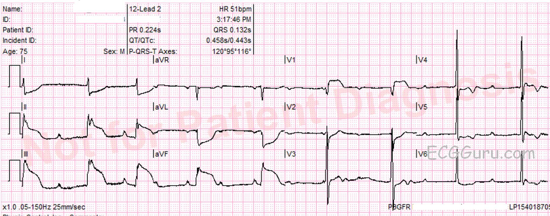
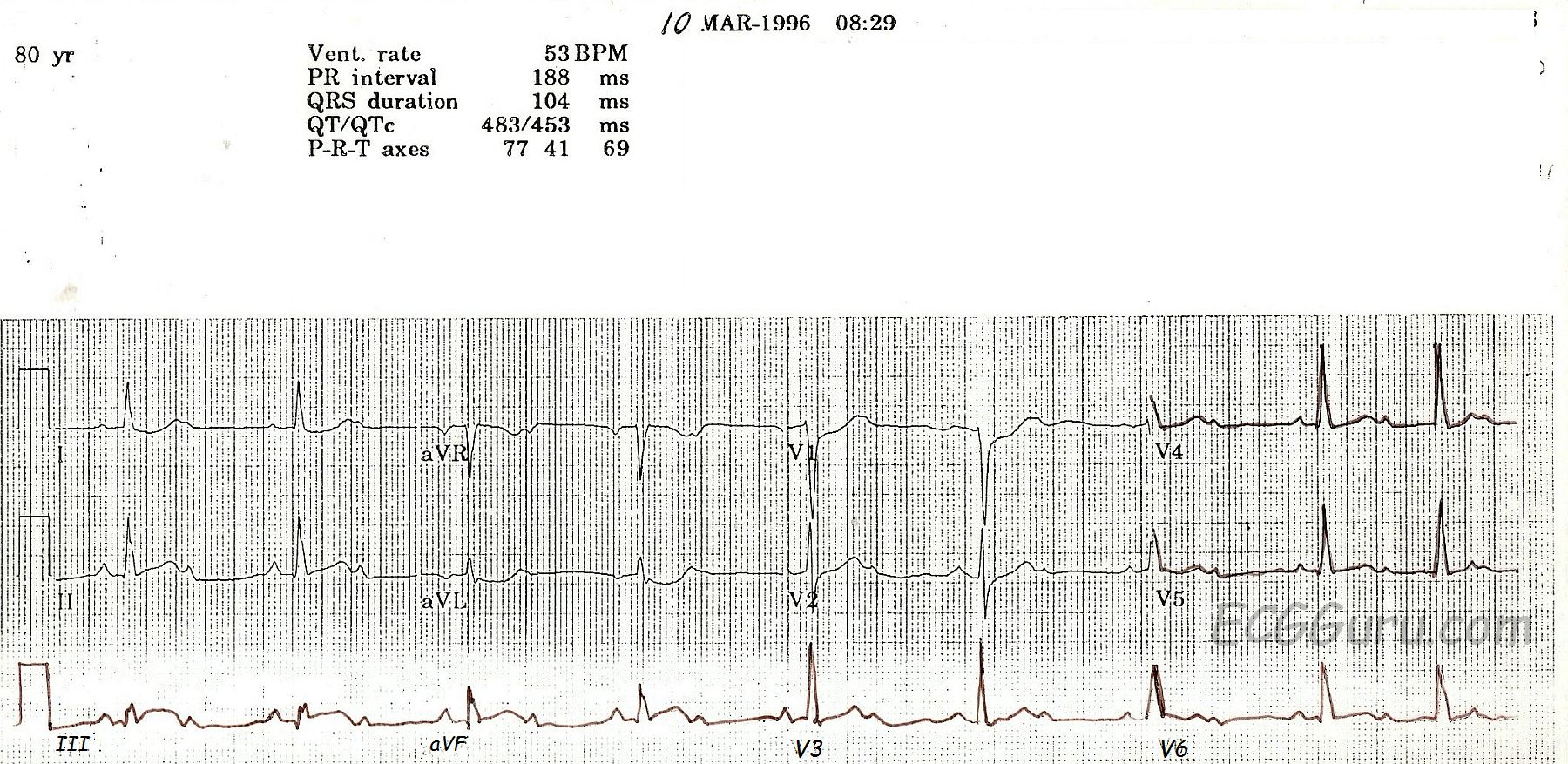
This ECG is from an 80-year-old woman who had an acute inferior wall M.I. with a second-degree AV block.
 
Some people incorrectly call ALL second-degree AV blocks that are conducting 2:1 "Type II".  This is incorrect, as Mobitz Type I can also conduct with a 2:1 ratio.  The progressive prolongation of the PR interval will not be seen with a 2:1 conduction ratio, because there are not two PR intervals in a row.

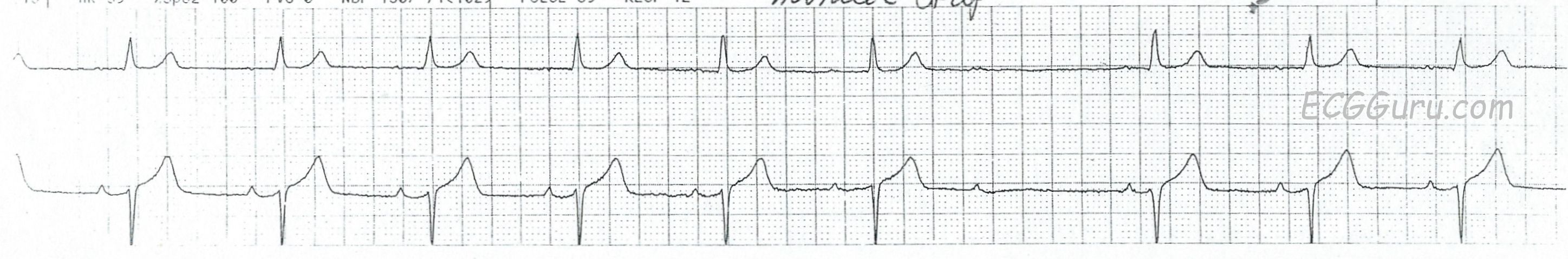
This is a good example of a Type I, or Wenckebach, block which is initially conducting 2:1.  At the end of the ECG, two consecutive p waves conduct, showing the "progressively-prolonging PR interval" hallmark of a Type I block. Type I blocks are supraHisian - at the level of the AV node - and generally not life-threatening.  Blocks that are conducting 2:1 present a danger, however, in the effect they have on the rate.  Whatever the underlying rhythm is, the 2:1 block will cut the rate in half!  This patient has an underlying sinus tachycardia at 106, so her block has caused a rate of 53.  In light of her acute M.I., that rate is probably preferable to the sinus tach. This patient’s BP remained stable, and she did not require pacing. 

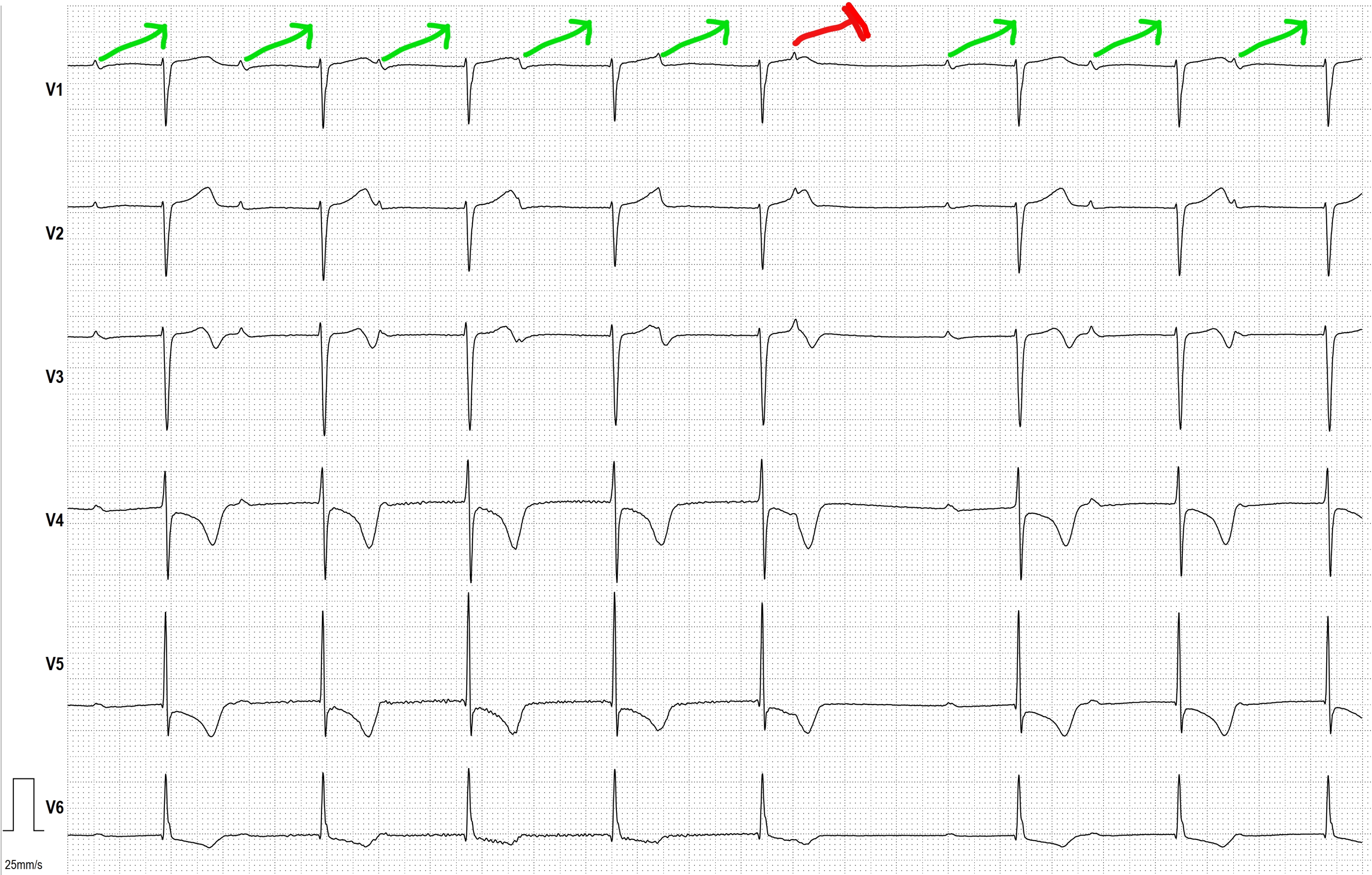
The ST signs of acute M.I. are rather subtle here. Note the "coving upward" shape in Lead III, and the reciprocal depressions in I, aVL, V1, and V2.  Type I blocks are common in inferior wall M.I., since the AV node and the inferior wall often share a blood supply - the right coronary artery. 

While the print quality of this ECG is not the best, it is a great teaching ECG because it starts out with 2:1 conduction, then at the end of the strip, proves itself to be a Wenckebach block.   

Dawn's picture

Second-degree AV Block, Type I

This 67 year old man is noted to have a slightly irregular pulse.  At the beginning of this ECG, he appears to be in NSR with a first-degree AV block.  Twice, P waves are non-conducted.  Careful measurement of the P to P interval shows that it is regular, there are no PACs noted.  The PR interval changes very subtly by lengthening just before the non-conducted P waves.  A hint when non-conducted P waves are noted, first check for non-conducted PACs.  If the sinus rhythm is regular, check the PR interval before the non-conducted beat, and the PR interval immediately after the non-conducted beat.  You will see the PRI preceding the non-conducted P is longer than the PRI after the NCP.

Wenckebach conduction is caused by RP/PR reciprocity.  In other words, the shorter the RP interval, the longer the PR interval.  So, as the PRI lengthens, the QRS "moves" to the right, eventually causing the next regular sinus P wave to fall into the refractory period and fail to conduct.  This results in a pause, or a long RP interval, which shortens the next PRI. 

 If you or your students would like to review AV Blocks, go to this LINK for Dr. Grauer's excellent, FREE, self-directed tutorial.

For a slightly more advanced discussion of RP/PR reciprocity, see Jason's Blog.


All our content is FREE & COPYRIGHT FREE for non-commercial use

Please be courteous and leave any watermark or author attribution on content you reproduce.