
Submitted by Dawn on Sat, 08/26/2023 - 16:53
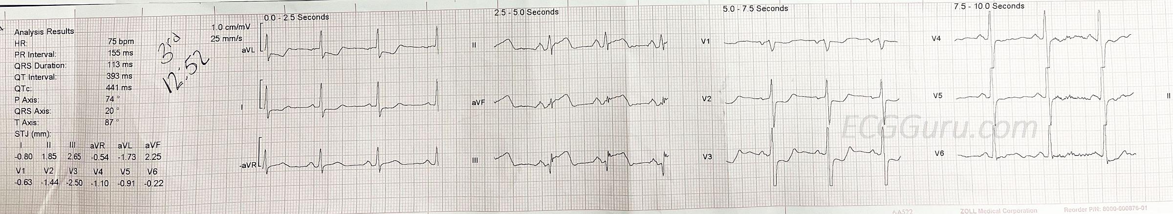
Does something about this ECG look "different" to you? This ECG shows a “classic” presentation of inferior-posterior M.I. when it is caused by a lesion in the right coronary artery (RCA). There are ST elevations in leads II, III, and aVF. Reciprocal ST depression is seen in Leads I and aVL. There is also reciprocal ST depression in Leads V1 – V3. These more rightward anterior leads are reciprocal to the posterior (or posterior-lateral) wall, so the ST elevation is actually posterior. Another sign that this is an RCA lesion is that the ST elevation in Lead III looks worse than the STE in Lead II. It would be helpful to check the right precordial leads, or at least V4 Right, as elevation there would indicate right ventricular M.I.
Depending on how experienced you are at evaluating ECGs, you might have immediately noticed something “different” about this tracing. It is printed in Cabrera format, which groups the leads (viewpoints) more geographically than a traditional ECG does. In addition to grouping the leads more geographically, instead of aVR, the machine records - aVR. That reverses the negative and positive poles of aVR, putting the positive ("seeking") electrode at 30 degrees - halfway between Leads I and II. Those of us who have been looking at ECGs for decades often feel a bit disconcerted by this format, because we have developed almost an intuitive way of seeing the ECG as a “map”, and this rearrangement thwarts our brains’ approach to the ECG. I would imagine, however, that this might make interpretation a bit easier for someone who is not prejudiced by the standard way of printing. This method is especially helpful when looking for inferior wall M.I., as we see here, because the lateral leads are together in a row, and the inferior leads are grouped together.
ECGs are “standardized” all over the world. This makes them easier for most of us to develop our interpretation skills. But, there are still options available to individuals, and some are popular in certain countries. Watch for Cabrera format, as well as changes in paper speed (50 mm/min or 12.5 mm/min instead of 25 mm/min). You might run across ECGs that have had the voltage measurements enhanced. You can determine this by looking at the standardization markers, which will be exactly 2 big boxes tall if set to the most common standard of 10 mm (10 small boxes) being equal to 1 mV.

Rate this content:
All our content is FREE & COPYRIGHT FREE for non-commercial use
Please be courteous and leave any watermark or author attribution on content you reproduce.
Comments
A Beautiful Example of Cabrera Format with Proximal RCA OMI
============
As an American-trained physician who practiced exclusively in the United States — I had not been exposed to ECG formats other than the standard format used in the United States. This changed for me in 2010 — at which time I began active participation in multiple ECG internet forums, many with a huge international audience. Even for those clinicians not participating in internet ECG forums — the internet has changed the world. Because of international travel — the chances are that all clinicians at one time or another will encounter in medical records or elsewhere, different ECG formats from countries other than their own. This occurs in today’s case — in which the Cabrera Format for limb lead sequencing is used.
For clarity in today’s tracing — I have increased the size of the lead labels — and outlined in red to emphasize that instead of lead aVR, the Cabrera Format uses a NEGATIVE Lead aVR!
I explain in detail the concept of the Cabrera Format in my ECG Blog #215 (GO TO — https://tinyurl.com/KG-Blog-215 ). In brief — lead sequencing with the Cabrera Format is MORE logical than the sequence used in most of the rest of the world. As per the insert I added below today’s tracing in my Figure — it can be seen that by using a +aVR Lead (which corresponds to a frontal plane axis of +30 degrees) — transition as one moves from the highest lateral lead aVL (at -30 degrees) — to the most rightward frontal plane lead ( = Lead III at +120 degrees) is gradual.
Isn’t it MUCH EASIER to appreciate the gradual transition in today’s tracing from marked reciprocal ST depression that we see in lead aVL — to maximum ST elevation in lead III?
NOTE #1: The Cabrera Format is used in several Scandinavian countries, in selected parts of Germany, and in selected other locations. You WILL on occasion run into this format IF you use the internet at all in your medical encounters.
NOTE #2: Most of the time — the Cabrera Format is accompanied by a faster recording speed (ie, of 50 rather than 25 mm/second). I discuss how to adapt to interpreting tracings at this different recording speed in my Blog #215.
NOTE #3: Today’s tracing strongly suggests acute proximal RCA (Right Coronary Artery) Occlusion because: i) There is an acute inferior STEMI with maximal ST elevation in lead III > II; ii) There is marked reciprocal ST depression in lead aVL; — and — iii) There is minimal (if any) ST depression in lead V1 — which suggests that ST elevation from associated acute RV MI is attenuating the amount of ST depression that we would expect to see in lead V1, in this patient who also shows evidence in leads V2,V3 of associated acute Posterior MI. Since the proximal RCA supplies the Right Ventricle’s vascularization — the diagnosis of probable associated acute RV MI in today’s case localizes the “culprit” artery to the proximal RCA (As per Dawn — obtaining right-sided leads would confirm acute RV MI).
NOTE #4: As I’ve frequently emphasized on the ECG Guru — there is NO need for doing posterior leads — as the positive Mirror Test for leads V2,V3 tells us there definitely is associated posterior MI.
====================
Ken Grauer, MD www.kg-ekgpress.com [email protected]