Dawn's picture

Today’s ECG of the WEEK comes from Sebastian Garay, Paramedic.  He presented it on his excellent website CardioCareConcepts.com, and was kind enough to share it with the ECG Guru.  It is a great example of LEFT MAIN CORONARY ARTERY lesion with ST elevation in aVR and V1.

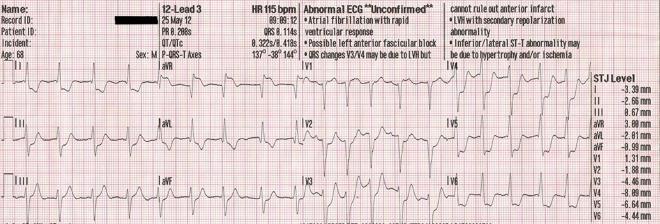
The patient was a 68 year old man who presented with a sudden onset of chest pain, followed by cardiac arrest.  He was revived by the use of an automatic external defibrillator (AED).  The initial 12-Lead ECG shows atrial fibrillation with a rapid response of 102 bpm.  There are prominent ST ELEVATIONS in aVR and somewhat more subtle STEs in V1.  These leads reflect the base of the septum, which is the area perfused by the proximal left coronary artery.  A lesion in this area is sometimes in the LEFT MAIN coronary artery, or both the proximal LCA and the circumflex.  Both of these types of lesions carry a very high mortality rate.

The widespread ST depressions reflect the injury current, which is being directed upward and toward the patient’s right shoulder, causing a reciprocal depression in all leads except aVR, V1 and Lead III.

This patient arrived in the Emergency Dept. in grave condition and was taken to the cath lab, where an occlusive lesion was found in the LEFT MAIN coronary artery.  He later died from this severe injury.

We recommend further reading on this topic, as there has been a large body of research on ECG findings of ST elevation in aVR.  Here are some links of interest:


Dr. Smith’s Blog;   JACC Online; ScienceDirect.com.

Rate this content: 
Average: 3.8 (5 votes)

Comments

ekgpress@mac.com's picture

 
     Our THANKS to Sebastian Garay for allowing the ECG Guru to publish this highly insightful tracing  obtained from a 68-year old man who presented with severe chest pain followed by cardiac arrest. Unfortunately  the patient succumbed from his extensive injury which was found on cath to be the result of severe LMain coronary disease. Details of the cardiac catheterization and of the timing of this ECG relative to this cath are unknown  so I will speculate.
 ---------------------------------------------------------------------
The ECG shows an irregularly irregular rhythm consistent with AFib (Atrial Fibrillation) with a relatively (but not excessively) rapid rate. The axis is leftward, consistent with LAHB = Left Anterior HemiBlock (predominantly negative QRS complex in the inferior leads). There is no chamber enlargement. The most remarkable finding relates to Q-R-S-T Changes:
  • A decidedly widened Q wave is present in lead aVL  with a smaller q in the other high lateral lead ( = lead I). There are also small q waves in lateral leads V5,V6.
  • Transition is delayed (the R wave becomes taller than the S wave only between V4-to-V5). That said  there appear to be small-but-present initial positive deflections in V1,V2 (albeit intermittently in V2 therefore no definitive indication of anterior infarction (LAHB itself may produce poor R wave progression with delayed transition due to resulting initial unopposed posterior forces that are seen with this hemiblock conduction defect).
  • There is dramatic ST depression in multiple leads. This suggests diffuse subendocardial myocardial ischemia. In contrast, there is ST elevation in “right-sided” ( = opposing) leads  especially in lead aVR (which shows 4mm ST elevation), but also in lead V1 and perhaps to a slight extent in lead III.
  • A final finding worthy of mention are disproportionately peaked T waves that are seen best in lead V3 (but perhaps also in V2,V4). While clearly not attaining the usual proportions of true DeWinter T wave status  there is J-point ST depression in lead V3 with rapid ascent of the T wave   so this picture that often characterizes impending LAD occlusion is at least suggested.
CONCLUSION: This ECG is not the picture of acute LMain occlusion! Despite cath and clinical findings that suggest this patient succumbed to massive infarction from LMain occlusion  the picture of diffuse ST depression with ST elevation in leads aVR and V1 with a hint of DeWinter T waves suggests significant coronary disease (ie, severe narrowing) that may well be due to impending occlusion in either the proximal LAD or LMain distribution. But NOT occlusion!  since with acute LMain occlusion the picture is one of diffuse ST elevation rather than diffuse ST depression.
  • MY SUSPICION: Given the unfortunate rapid demise of this patient  I suspect that this ECG was obtained on ED arrival PRIOR TO occlusion of the LMain coronary artery  with rapid deterioration following shortly after occlusion took place ...

---------------------------------------------------------------------

Please CHECK OUT this pdf on Clinical Use of Lead aVR in ECG Interpretation (excerpted from my ECG-2014.ePub). GO TO http://tinyurl.com/ECG-9-aVR 
  • The part relating to use of aVR for assessment of severe coronary disease begins in Section 09.40 of the pdf.
 ---------------------------------------------------------------------

Ken Grauer, MD  www.kg-ekgpress.com   [email protected] 

All our content is FREE & COPYRIGHT FREE for non-commercial use

Please be courteous and leave any watermark or author attribution on content you reproduce.