Dawn's picture


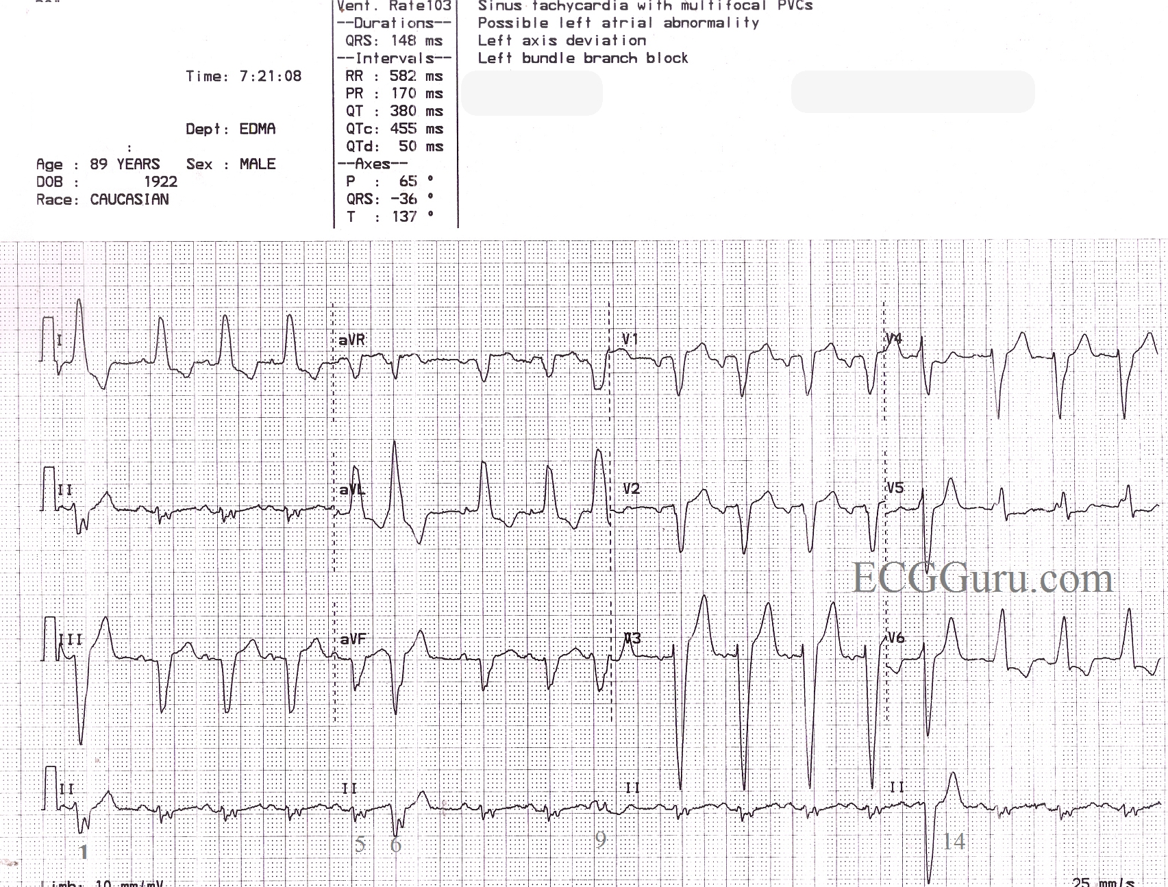
This is a great ECG for teaching your students about some of the different causes of wide QRS.  This 89 year old man has a sinus rhythm that is around 100 bpm, and his QRS is widened at 148 ms (.148 sec).  Leads I and V6 are positive, and Lead V1 is negative, meeting the criteria for left bundle branch block. There is a left axis deviation, which is common with LBBB, although it is not always this pronounced, indicating that there is possibly another cause for LAD.  In this ECG, there are also PVCs and probable fusion beats.  The 14th beat is a PVC.  Complexes 1, 6, and 9 are possibly fusion beats. Fusion can be described as an almost simultaneous sinus beat and ventricular beat.  The depolarization waves, one coming from the top of the heart and one coming from the bottom, meet and "fuse" on the ECG.  Fusion beats will have some characteristics of the supraventricular beats and some of the ventricular beats.  They are not significant except that fusion can be said to "prove" the existence of a ventricular pacemaker - either a natural pacemaker or an electronic one.

Do you see anything else interesting in this ECG?  How would YOU describe this rhythm?  Please do not hesitate to add your comments, or ask questions of the experts who contribute to this site.  We will respond quickly to all questions.

Rate this content: 
Average: 3.7 (3 votes)

Comments

ekgpress@mac.com's picture

This is LBBB - plus a LOT more (the patient has had 89 years to get to this point - and it shows .... ). There are BASIC Teaching Points and many ADVANCED Teaching Points illustrated by this tracing which I'll comment on below.

The RHYTHM:

  • Underlying sinus rhythm as per Dawn.
  • Erratic baseline with artifact that makes some assessment of rhythm quite difficult ....
  • Starting with what we KNOW - Beat #14 does look like a PVC (albeit baseline artifact before this QRS prevents me from being certain that no premature P wave is present ... ).
  • I'm not sure what beat #9 is. Although it initially looks in the lead II rhythm strip at the bottom like there is a premature P wave = PAC (slightly early atrial beat that looks different from other P waves) - in simultaneously occurring leads aVR,aVL,aVF - it seems this deflection that looks like a PAC in lead II is actually part of the QRS complex - with this QRS looking different than the "normal" LBBB-conducting QRS (though I think it not necessarily a fusion beat).
  • Beat #16 seems to occur a tad early with a QRS that looks like the normal LBBB-conducting QRS - so perhaps this is a PAC (too much baseline artifact preceding this beat to be sure ... ).
  • It almost looks like a different P wave precedes beat #1 - so whether this is a fusion beat vs PAC with aberrant conduction (on top of the LBBB) I think is uncertain ....

BOTTOM LINE re RHYTHM - It is not easy to be sure of what is going on. In situations like this - I find it best to state the generalities we can be sure of which is: Underlying sinus rhythm - multiform PVCs - possible PACs - there may be some fusion complexes.
-----------------------------

As to the REST of the ECG:

  • LBBB (as per Dawn - with typical monophasic R wave in leads I,V6 and negative QRS in lead V1).
  • LAE (left atrial enlargement) - seen from the very deep and rounded negative P in lead V1.
  • Probable LVH (seen from the very deep >25-30mm S wave in lead V3 - which with LAE makes LVH >90-95% likely in a patient with LBBB).
  • Secondary ST-T wave changes in leads V5,V6. It is always a challenge to assess ST-T wave changes in the setting of BBB - because the BBB itself will cause changes. One expects that the ST-T should be oppositely directed to the last QRS deflection in the 3 key leads (I,V1,V6) - which it is here. But lead V5 shows J-point ST depression and ST flattening - and - the J-point in lead V6 is down >3mm - and that is a bit unexpected for typical LBBB - so I'd suspect ischemia (though ST-T wave changes do not really appear to be new ... ).

-----------------------------

OVERALL IMPRESSION:

  • Sinus rhythm - multiform PVCs - possible PACs
  • LBBB
  • LAE/probable LVH - Patient likely has cardiomyopathy (given patient's age and presence of LAE/probable LVH/LBBB/multiform PVCs).
  • Secondary ST-T changes consistent with ischemia - though probably no acute change. Suggest clinical correlation. Not knowing the clinical situation - one wonders if it might not be best to leave this man who has made it to 89 despite all of the above changes alone .... (but he might have clinical heart failure that can be treated - so the History will "tell the tale" of what is best to do).

-----------------------------

For anyone wth an interest - LINKS to:


Ken Grauer, MD  www.kg-ekgpress.com   [email protected] 

All our content is FREE & COPYRIGHT FREE for non-commercial use

Please be courteous and leave any watermark or author attribution on content you reproduce.