Displaying 1 - 9 of 9
Dawn's picture

Widespread ST Elevation With Right Bundle Branch Block

Usually, instructors of basic ECG classes look for examples of the most common conditions that are likely to be encountered by the learners.  But, sometimes, it is advantageous to show students more unusual presentations to remind them of the infinite possibilities when we care for living beings.  This series is a very good example of what can and does happen to some people with cardiovascular disease.  It will give your students an opportunity to think about possible interpretations, and also about anticipating clinical implications and emergencies that may arise.

The Patient:  This patient is a man in his 80s who has been active his whole life.  He considers himself to be healthy, giving no medical history and denying medication use. He states that he has had a yearly health exam.  Today, he felt “tired and dizzy” while raking leaves.  As he walked to his house to rest, he had a syncopal episode and fell, hitting his head. He was unconscious for a few minutes. A family member called for Emergency Medical Services (EMS). Paramedics found him awake and complaining of bilateral “shoulder and wrist” pain. He had no obvious trauma to his extremities, but had some bruising on his head and face.  He denied recent illness and substance abuse.  He was oriented x3. He was pale and diaphoretic, and complained of nausea. He denied chest or back pain.  He denied shortness of breath.  BP 100/60.  Heart rate bradycardic.  SPO2 above 95%.  He was given aspirin and ondasetron, and transported to a hospital.

Dawn's picture

Inferior Wall, Posterior Wall, and Right Ventricular M.I.

The patient:    79-year-old man complaining of severe “burning” chest pain, radiating to his neck. Walking exacerbates his discomfort.  He has had nausea and vomiting for 24 hours. Past medical Hx includes high cholesterol and atrial fibrillation. Medications not known.


The ECGs:  These ECGs could be called “classic”.  There is a 100% occlusion of the right coronary artery (RCA), which was successfully repaired in the cath lab.  About 80% of inferior wall M.I.s are due to occlusion of the right coronary artery.  Depending on how proximal the occlusion is, we can expect a pattern on the ECG representing injury to all areas supplied by the RCA.  This “package deal” can include:

·         Inferior wall ST elevation.

·         Posterior wall extension.

Dawn's picture

Inferior Wall M.I. With Atrial Fibrillation or Atrial Flutter

The Patient   This ECG was obtained from a 74-year-old man who had a history of COPD. He was complaining of severe chest pain at the time of the ECG.

The ECG     The rhythm is atrial fib or flutter (the R to R intervals are irregular, but seem to repeat about 4 interals).  Flutter waves are seen during some of the longer intervals. The rate is approximately 90 beats per minute.  The ST segments are very noticeably elevated in Leads II, III, and aVF.  There is reciprocal ST depression in Leads I and aVL, and also in all the precordial leads. 

Dawn's picture

Inferior Wall M.I. and Right Bundle Branch Block

These ECGs were taken from a 76 year-old-man who was complaining of chest pressure for 20 minutes.  He had a remote history of coronary artery bypass graft surgery.

This case has several good teaching points, including:

Significant artifact.  The limb leads show artifact which is severe enough to hamper our assessment of the j point location. Every effort should be made to eliminate artifact.  Some measures that might help are:

        *  clean and slightly "rough up" the skin where the electrode will be placed.  A rough wash cloth or gauze pad will work.

        *  shave hair if necessary.

        *  avoid areas of movement if possible.  Precordial electrodes must be placed in specific spots, but limb leads may be placed anywhere on the limb or on the trunk if it is impossible to avoid movement on the limbs.

       *   use fresh electrodes that have been protected from drying out.

Subtle STEMI changes.   This patient has an inferior wall M.I., which was confirmed as a complete occlusion of the right coronary artery in the cath lab.  The ST elevation in Leads II, III, and aVF is subtle, and more difficult to measure because of the artifact.  However, the SHAPE of the ST segments is a giveaway - they are very straight.  A convex-upward shape is normal (see Lead I).  Also, Lead aVL shows typical ST DEPRESSION, as a reciprocal view of the STE in Lead III.  More ST depressions can be seen in Leads V1 through V3, and they end abruptly there.  These localized ST depressions represent a reciprocal view of the posterior (also called lateral) wall, and represent an "extension" of the inferior wall M.I. up the back of the heart.  A V4 Right lead was obtained and shows no measurable ST elevation, but the shape is straight to slightly "frowning", indicating that the right ventricle may soon have STE.  Repeat ECGs should be obtained to watch for more definite ST elevations.

Dawn's picture

Circumflex Occlusion with Posterior-lateral M.I.

This ECG was obtained from a woman with chest pain who was taken to the cath lab and found to have a 100% occlusion of her circumflex artery.  

There are obvious ST segment elevations in Leads I and aVL, as well as in Lead II.  Lead II is the most leftward of the inferior wall leads, and I and aVL reflect the high lateral wall. She also has ST depressions in V1 through V3.  If you look closely at the R wave progression in the anterior leads, you will readily note that it appears that V1 and V3 wires have been reversed.  That being said, the "real" V2 and V3 have taller-than-normal R waves.  The tall R waves and ST depression are signs of "posterior wall M.I."  Recently, the actual definitions of the "lateral" wall and "posterior" wall have come into question.  However, the important thing clinically, is that this patient IS experiencing an ST elevation M.I. (STEMI), which was confirmed in the cath lab.  The locations of the ST changes were consistent with the 100% occlusion of her circumflex artery.

For our more advanced readers (and our "Gurus"), there is an interesting rhythm.  The P wave morphology changes frequently, even though the rhythm remains regular.  The rate, at 62 BPM, was adequate, and the patient did not suffer any consequential dysrhythmias during her procedure.  We don't have long-term followup information on her.


Dawn's picture

Inferior-lateral and Posterior Wall M.I.

This is from a Cardiac Alert patient, with chest pain, in the Emergency Department.  The ECG shows ST elevation in the inferior leads (II, III, and aVF), and in the low lateral leads (V5 and V6).  There is reciprocal depression in V1 and V2, indicating injury in the posterior wall.  One could argue that "inferior" is just the term we use for the lower part of the posterior wall - the part that faces the floor in a standing person.  So, "inferior-posterior" reflects a more proximal occlusion of the culprit artery.

The high lateral wall is represented by I and aVL.  These leads would usually show marked reciprocal ST depression when II, III, and aVF have elevation.  However, in this ECG, aVL is depressed, but not as much as expected, and Lead I almost looks elevated!  This could represent even more extensive lateral wall involvement.  A dominant right coronary artery could be the culprit, but it seems more likely that a dominant circumflex artery is to blame, as it could perfuse the entire lateral wall before joining with the posterior descending artery and perfusing the inferior wall.  Unfortunately, we do not have the cath results on this patient.

The ST elevation in this ECG has the classic appearance of acute M.I., and will be interesting to both beginner and advanced students.

Often, one ECG can provide a wealth of teaching opportunities, no matter what the level of your students.  For the student learning to monitor the rate and rhythm, you might crop this image to only show the Lead II rhythm strip at the bottom, for a good example of normal sinus rhythm with a borderline PRI of .20 sec.   For the student learning about ST elevation M.I., this is a good example of inferior-posterior and lateral injury.  Leads aVL, V1 and V2 demonstrate reciprocal ST depression.  When an observant student notices the slight ST elevation in V6, a discussion of coronary artery distribution can occur.  

Dawn's picture

Artifact on an ECG Showing Inferior, Posterior, Lateral M.I.

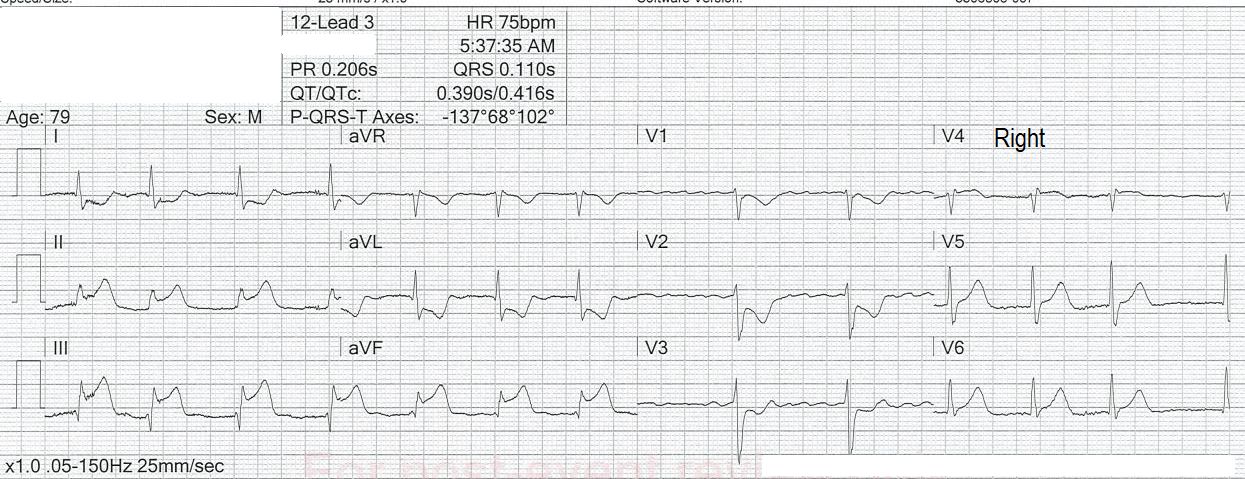
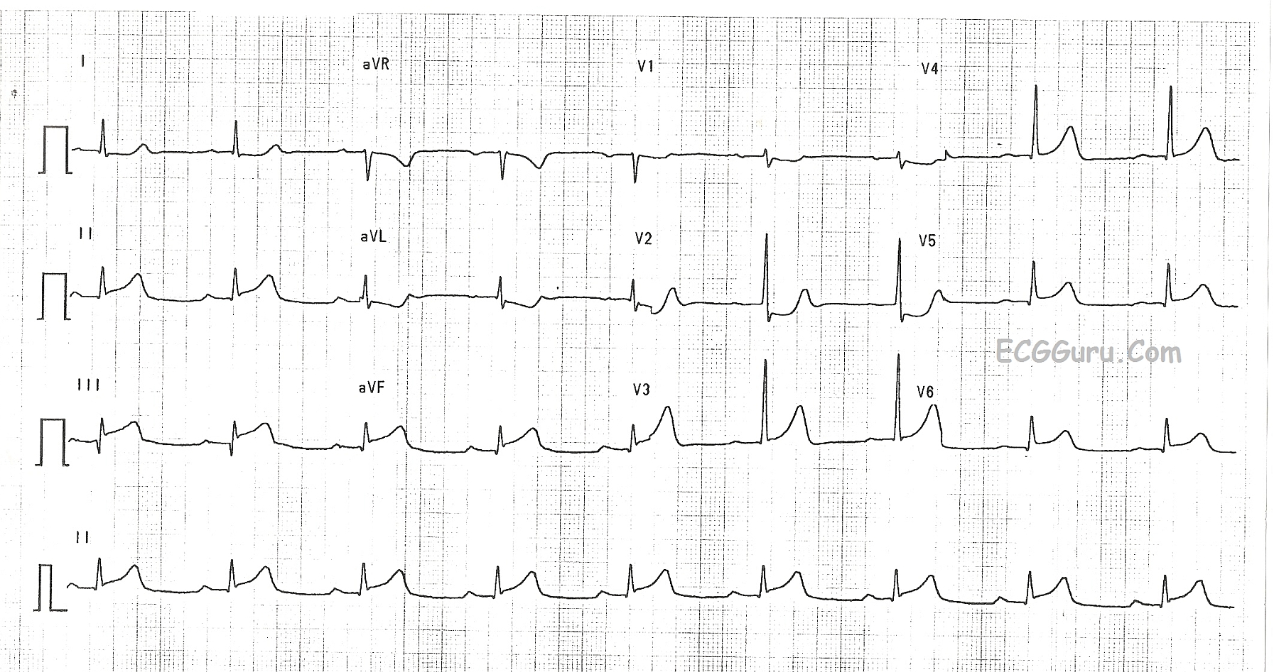
If you are an ECG instructor, it is important that you address the subject of artifact on the ECG.  Artifact has many causes, and it is important eliminate it whenever possible.  We should strive for the "cleanest" ECG possible.  As you can see in this example, the presence of artifact has caused the machine's computer rhythm interpretation to be incorrect.  The noisy baseline has caused the computer to call this rhythm "atrial fibrillation", but we clearly see P waves in all leads, especially in Lead II.  We recognize these P waves as authentic because they are regular, they  all look alike, and they have the same relationship to the QRS complexes each cycle (PR interval is the same).  

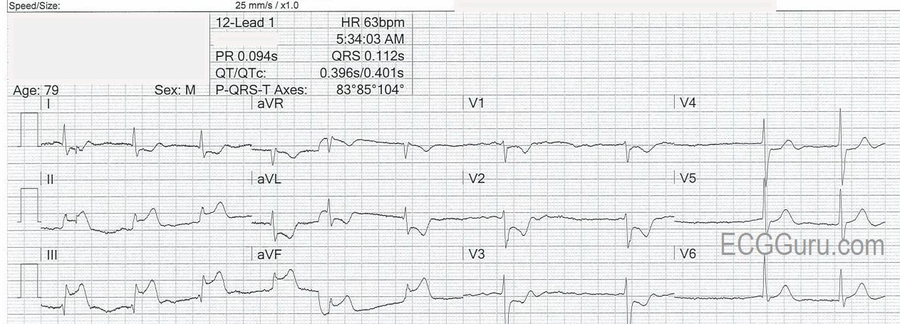
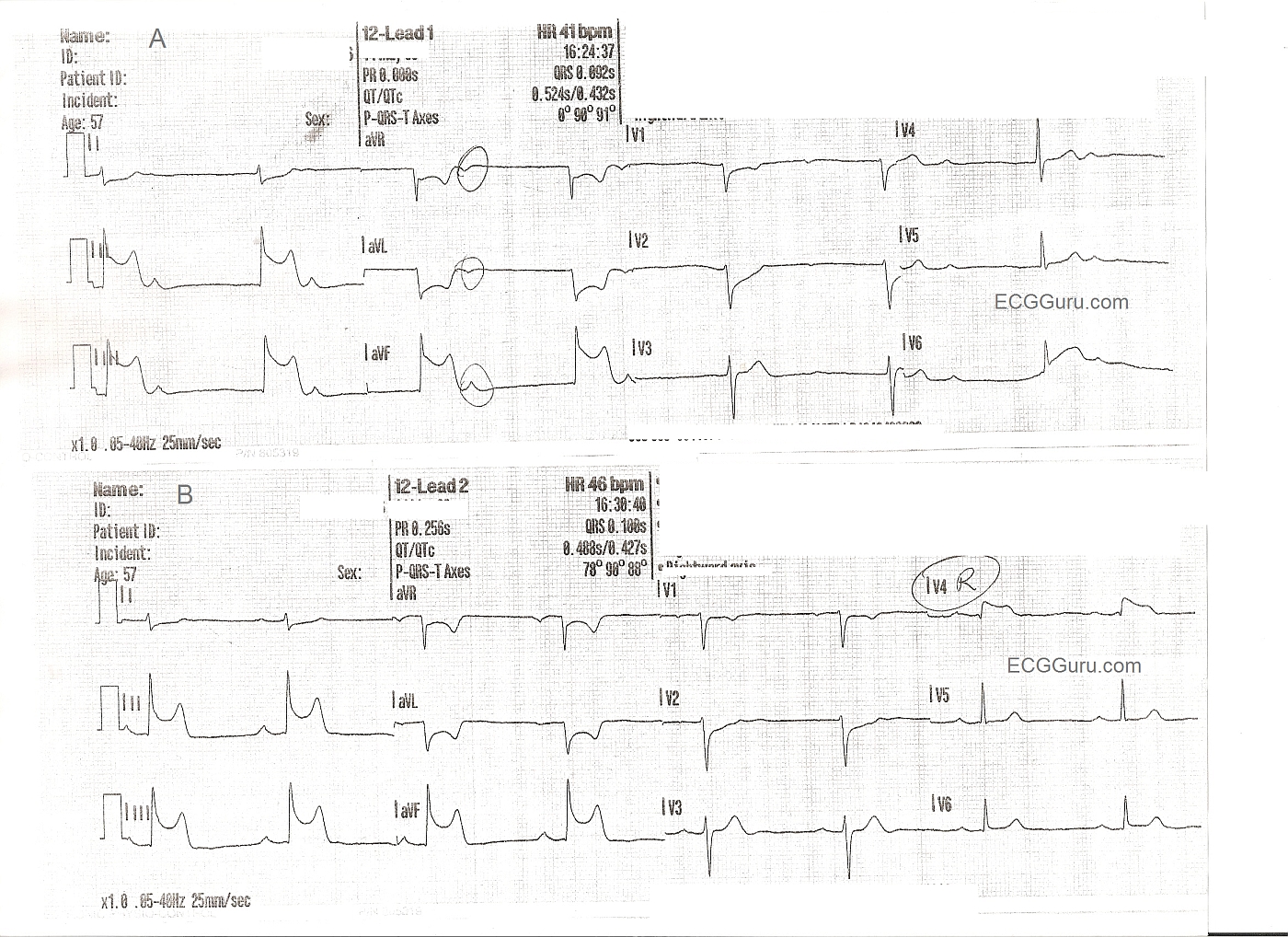
The patient is suffering a very large M.I., showing as ST segment elevation in Leads II, III, aVF, with slight elevation in V5 and V6.  In addition, Leads V1 through V3 have definite  ST depression, indicating extension of the inferior wall injury up the posterior wall of the heart.  There has been quite a bit of discussion lately in the literature about whether to call this a "posterior" M.I, or "high lateral", or just "inferior".  Semantics aside, the involvement of so many leads tells us that this  is a large M.I.  The patient was in the Emergency Dept. complaining of chest pain.

It is fortunate that the artifact did not affect our ability to see the ST elevation, but it could have.  And, of course, we would not want to treat this patient's "atrial fib" based on the machine interpretation.  But, it is always prudent to try to get rid of artifact.  In this example, Lead III has no artifact, so it could be assumed that the right arm electrode is the culprit, as Lead III does not utilize the RA electrode, and the other leads do.  

Troubleshoot for the cause of the artifact, and then retake the ECG.  Some common causes of baseline  artifact of this nature include:  patient movement, loose electrode, dried electrode, something touching the electrode, faulty or broken lead wire, and poor skin contact due to substances on the skin.  The electrodes should be fresh from the package, and applied to skin that is clean and dry.  The patient should be encouraged to relax and hold still (not so easy for a patient in distress).  Others at the bedside should avoid touching or manipulating the limbs of the patient during acquisition of the ECG data.  This only takes about 10 seconds.  I have seen artifact many times when a patient's blood was being drawn during the ECG, and the patient was squeezing his fist for the phlebotomist.

Dawn's picture

Inferior - Posterior M.I.

This ECG shows a classic inferior - posterior STEMI.  This M.I. was due to complete occlusion of the right coronary artery.  ST elevation apparent in Leads II, III, and aVF show the acute injury in the inferior wall, while ST depressions in V1 and V2 are reciprocal of the ST elevations in the posterior wall.  The tall R waves in Leads V1 - V3 most likely are reciprocal to pathological Q waves in the posterior wall.  Tall R waves in the right precordial leads can be caused by other cardiac conditions, such as right ventricular enlargement.  RV hypertrophy can probably be ruled out in this case because there is no right axis deviation or P pulmonale.  Because inferior wall M.I.s often extend into the posterior wall, it is the most likely cause of the tall R waves.

Dawn's picture

Inferior Wall M.I. with Right Ventricular M.I.

These two ECGs are from a 57 year old man with chest pain. The initial ECG shows ST elevation in Leads II, III, and aVF - inferior wall STEMI. Reciprocal changes are as expected in I and aVL. Reciprocal ST depression also seen in V1 and V2 indicate probable posterior wall involvement. Not surprising since the inferior wall is simply the lower part of the posterior wall. The first ECG also shows the patient in sinus brady with junctional escape: AV dissociation. The sinus node is often affected in IWMI that is caused by right coronary artery occlusion. The second ECG shows a slight increase in the sinus rate, and a sinus bradycardia. A V4 right lead has been performed, clearly showing ST elevation, and indicating right ventricular M.I.


FOR MORE INFORMATION ABOUT RIGHT VENTRICULAR M.I.,

Click Here

And Here

ECG Guru Resources

All our content is FREE & COPYRIGHT FREE for non-commercial use

Please be courteous and leave any watermark or author attribution on content you reproduce.