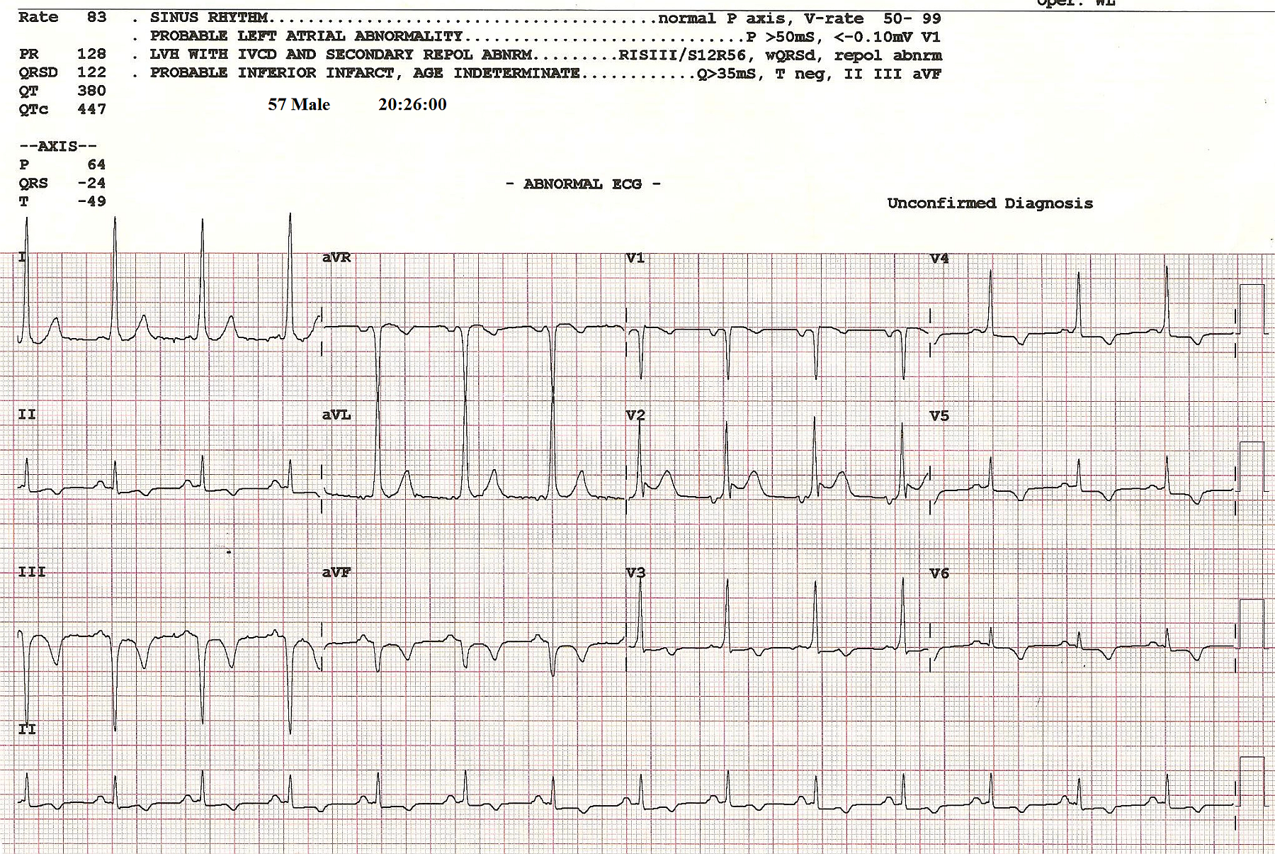
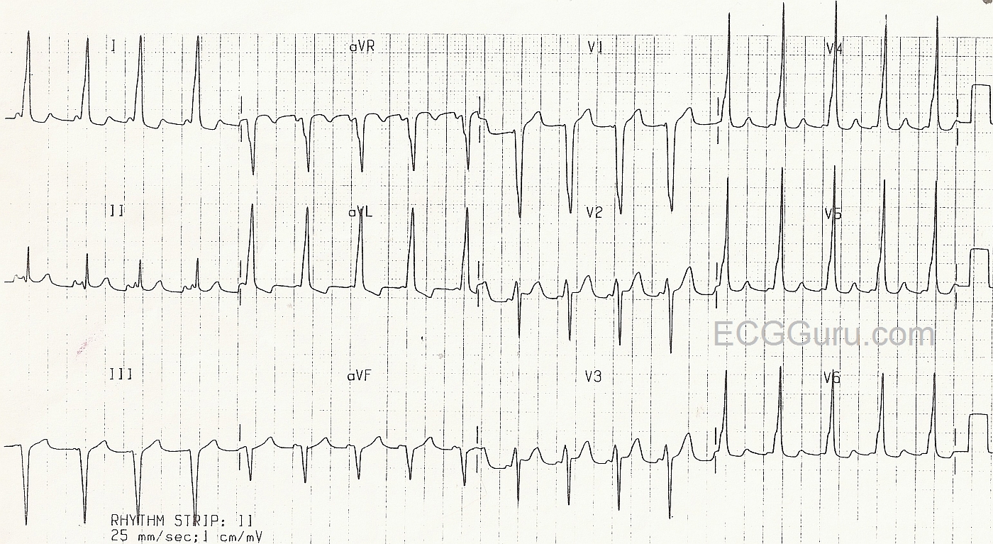
This ECG was submitted by Sebastian Garay, EMT-P and ECG Guru (and ECG Guru Member sebmedic). It is a very interesting case of wide complex tachycardia in a patient with Wolff-Parkinson-White.
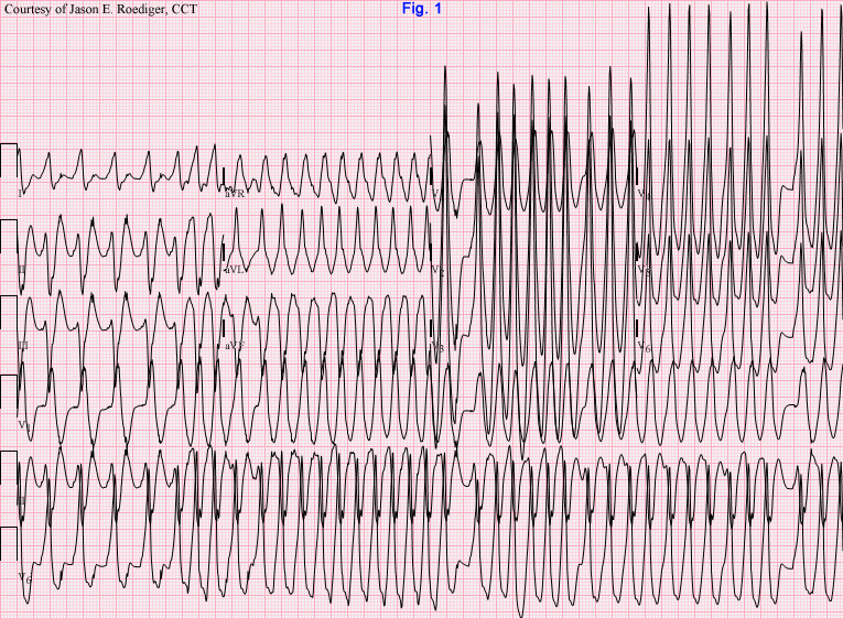
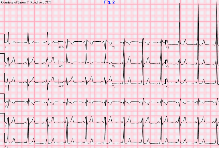
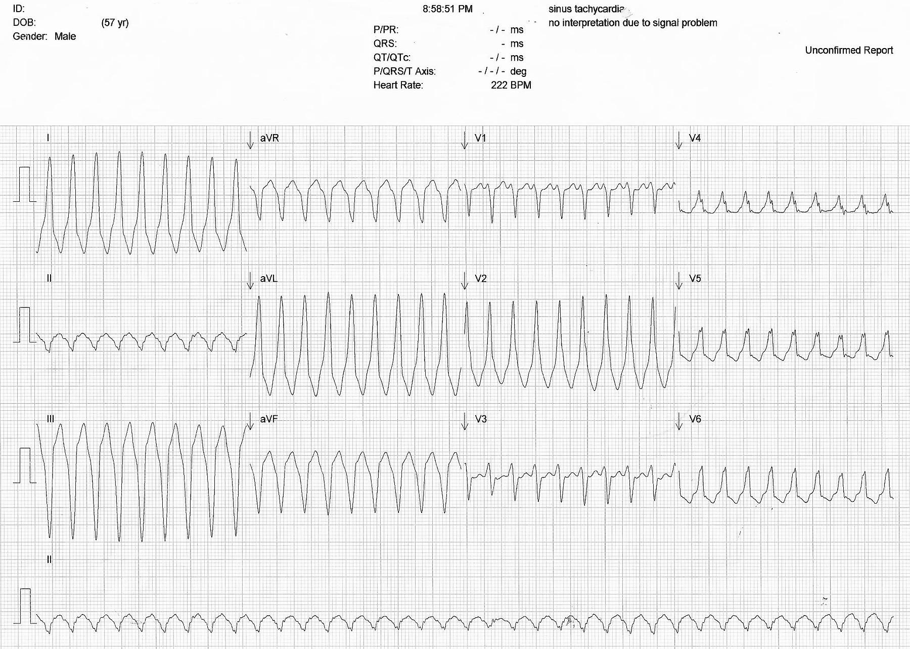
This is from a 57 year old man who sought medical help for a complaint of palpitations. He was known to have WPW. The paramedics determined that he was hemodynamically unstable, and in light of the wide-complex tachycardia, they performed a synchronized cardioversion at 100 j. The patient converted to the rhythm shown in the next ECG. The paramedics then administered a Lidocaine bolus and drip, considering this to be a ventricular tachycardia. The patient arrived in the Emergency Dept in improved hemodynamic condition. The first ECG shows a wide-complex tachycardia, and the second shows NRS with delta waves, indicating Wolff-Parkinson-White Syndrome.
Wolff-Parkinson-White syndrome involves an accessory pathway that allows atrial impulses to go around the AV node, entering the ventricles early. When the accessory pathway connects to the normal pathway through the AV node, a circle forms. This can lead to re-entry and fast rhythms. Conduction over the accessory pathway "pre-excites" the ventricles, causing a delta wave on the ECG. When the impulse enters the ventricles through the accessory pathway and then travels back up to the atrial using the normal AV node pathways, a wide-complex tachycardia is produced. The delta wave is what makes the QRS wide. On the other hand, when the impulse travels down the normal pathways and up the accessory pathway, a narrow-complex tachycardia will be produced.
The differential diagnosis here is 1) V Tach and 2) SVT with aberrant conduction or left ventricular conduction delay. 3) SVT due to re-entry through an accessory pathway. The second, post-conversion, ECG proves that this is W-P-W.