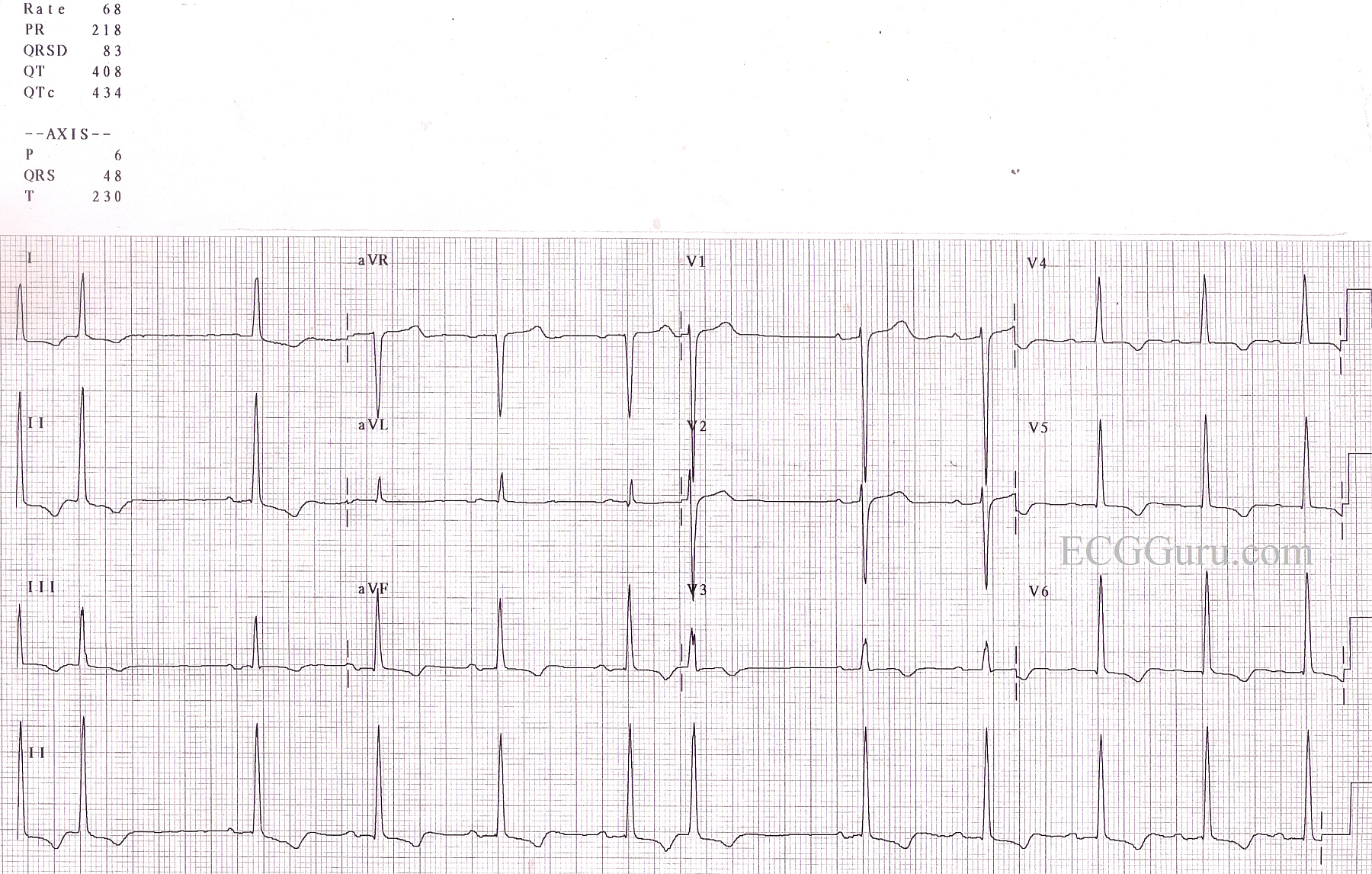
This ECG is from an elderly man with long-standing hypertension. It is a good example of LVH, with tall QRS complexes in the left-sided leads (V5, V6) and deep QRSs in right sided chest leads V1 and V2), but a rather unusual axis, in that it is normal, and we often seen left axis deviation with LVH. His hypertrophy is not severe as seen on imaging studies, but he shows an obvious "strain" pattern of sloping ST depression and T wave inversion in leads with upright QRS complexes. The signs of LVH are subtle, but when viewed as a whole ECG, the pattern seems more obvious. Always consider body shape, also, as a very thin person may produce more voltage on the ECG since the electrodes are close to the heart, and a person with a large chest may seem to minimize the QRSs, as we sometimes see with COPD. For more discussion of LVH criteria, go to the "Favorites" page. Every ECG blogger on the blogs list will have a discussion of LVH criteria and significance.
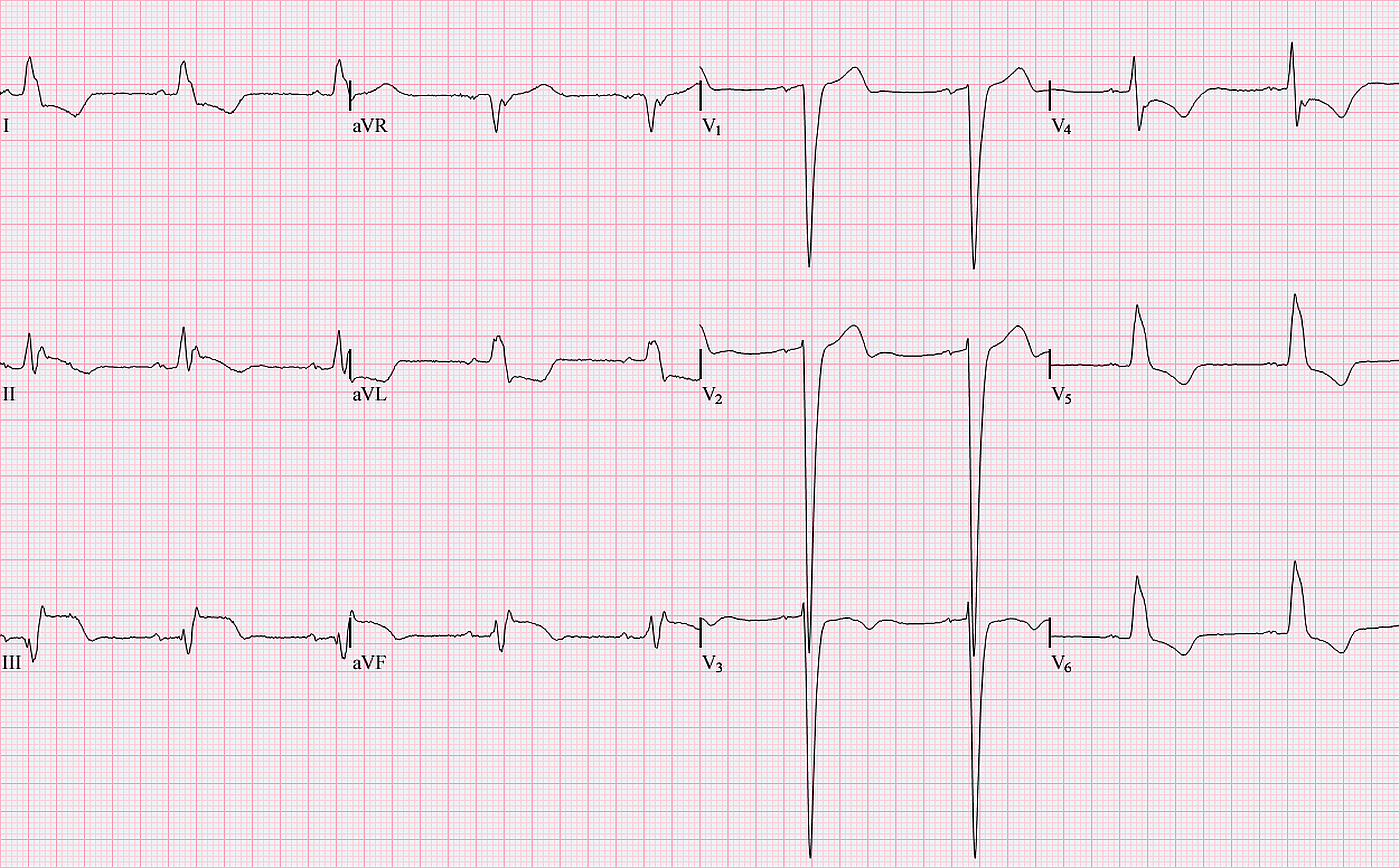
This interesting and instructive ECG was contributed by Jason Roediger, ECG Guru Extraordinaire, and one of the experts featured on our '"Ask the Experts" page. It is an excellent example of acute inferior wall M.I. with left bundle branch block. Left bundle branch block normally displays ST elevation and depression in a "negative concordance" pattern. That is, when the QRS complex is negative, we may expect ST elevation. When the QRS is positive, ST depression is seen. In this ECG, there is clearly ST elevation in Leads II, III, and aVF, and the ST segments have a distinct coved upward appearance. This shape signals to the experienced ECG interpreter that there is an acute injury. This ECG also shows voltage criteria for left ventricular hypertrophy, seen in the right chest leads, but not the left.
Unfortunately, the normal deviations of the ST segment seen in left bundle branch block can make diagnosis of acute M.I. difficult. For a good example of a left BBB without acute M.I., please refer to the ECG archives on this site. Often, students are taught that it is IMPOSSIBLE to see an acute M.I. in the presence of LBBB. This is not true, as this ECG clearly illustrates. See the March 4, 2012 blog post on the ECG Guru regarding this topic.