This ECG was obtained from a 37-year-old male who was complaining of non-radiating substernal chest pain.He offered no significant medical history.He denied taking any medications.He was hypertensive and bradycardic on arrival in the Emergency Dept. He was alert and ambulatory.Approximately 20 minutes after first being seen by paramedics, he suffered an episode of ventricular fibrillation in the E.D.He was resuscitated and sent to the cath lab.His coronary arteries were without lesions.We do not know the results of any lab tests, including troponins.
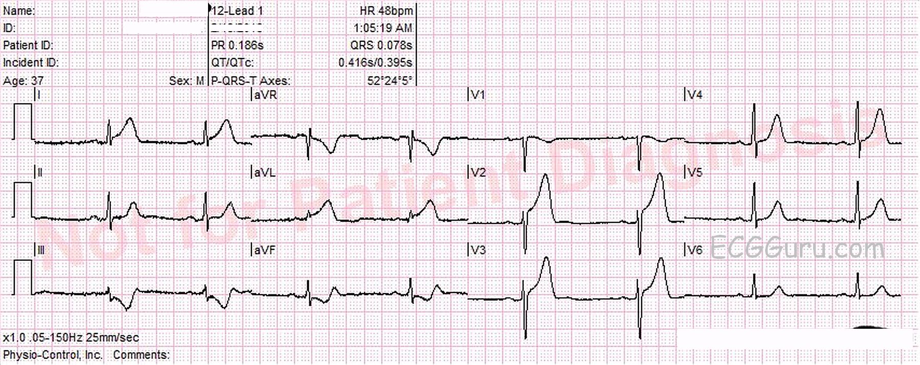
What does the ECG show?The rhythm is sinus bradycardia at a rate of 48 bpm. The PR, QTc, intervals and QRS duration are normal. The QRS frontal plane axis is normal and there is good R wave progression in the precordial leads.There is ST segment elevation in Leads I, aVL, V2, and V3, with reciprocal ST depression in Leads III and aVF.The ST segments that are elevated retain a relatively “normal” shape, being concave upward. There are no abnormal T wave inversions or pathological Q waves.