Submitted by Dr A Röschl on Sun, 09/10/2023 - 08:05
A common cause of pauses and bradycardia are non-conducted PACs, which generally do not require treatment. Therefore, it is important to differentiate between pauses or bradycardia that require treatment.
Submitted by Dr A Röschl on Sat, 06/03/2023 - 04:32
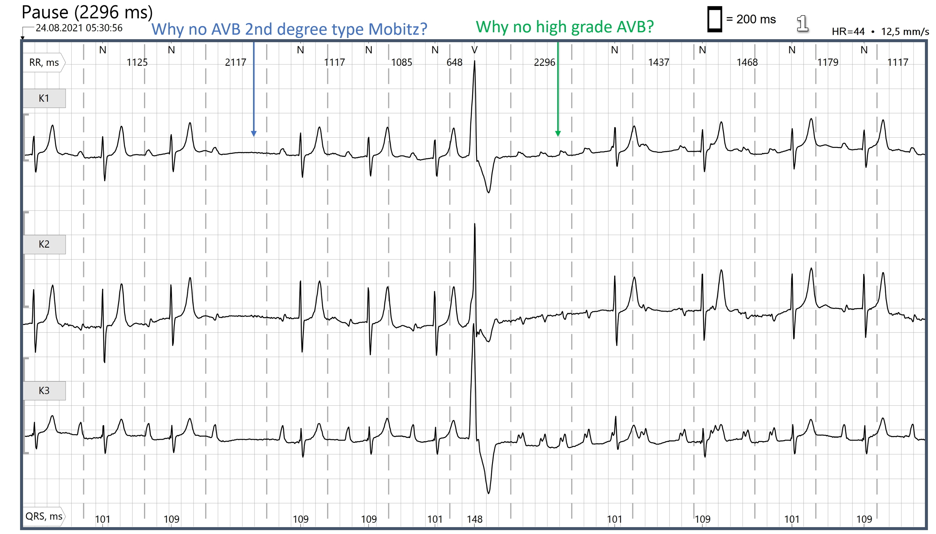
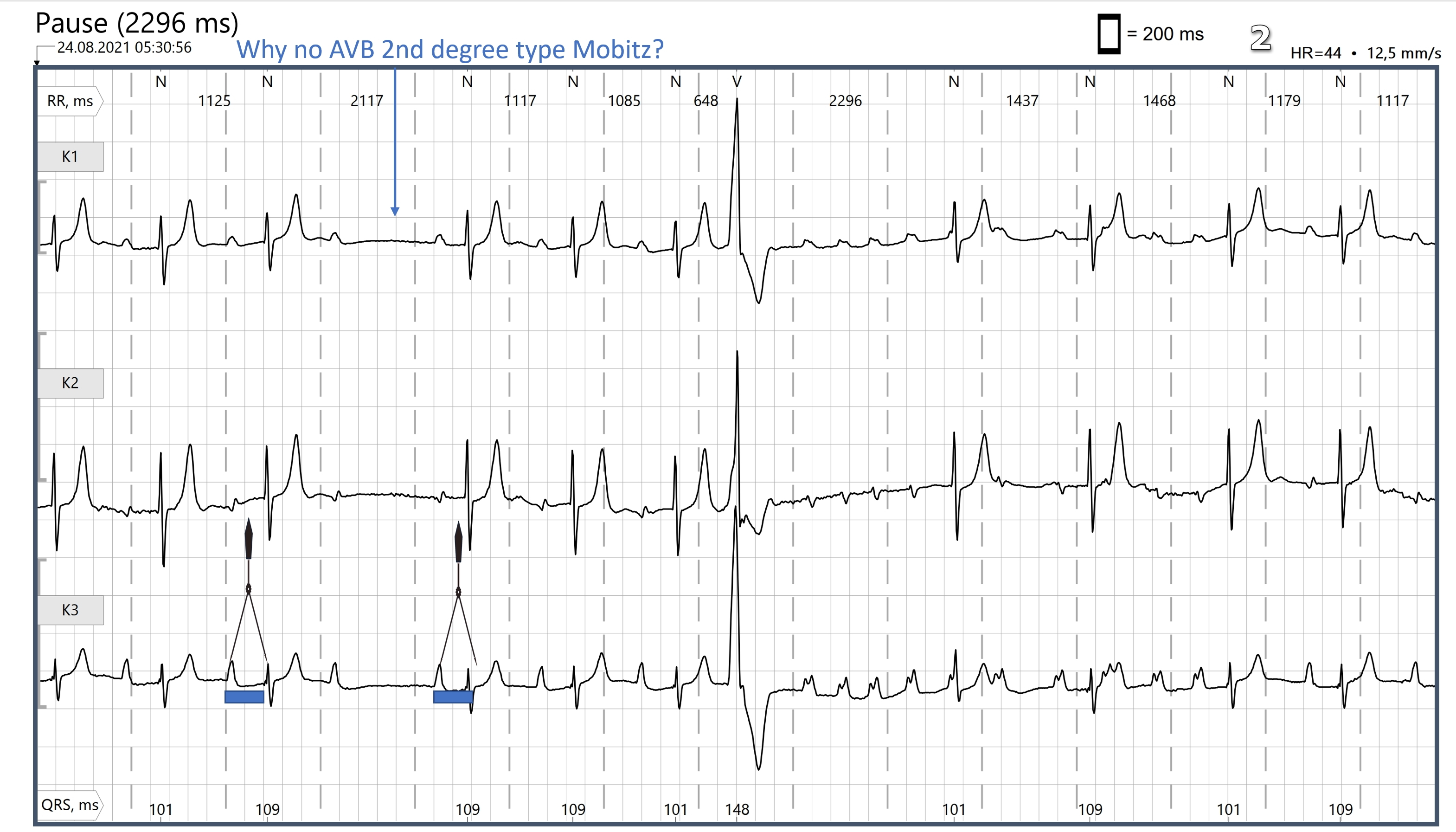
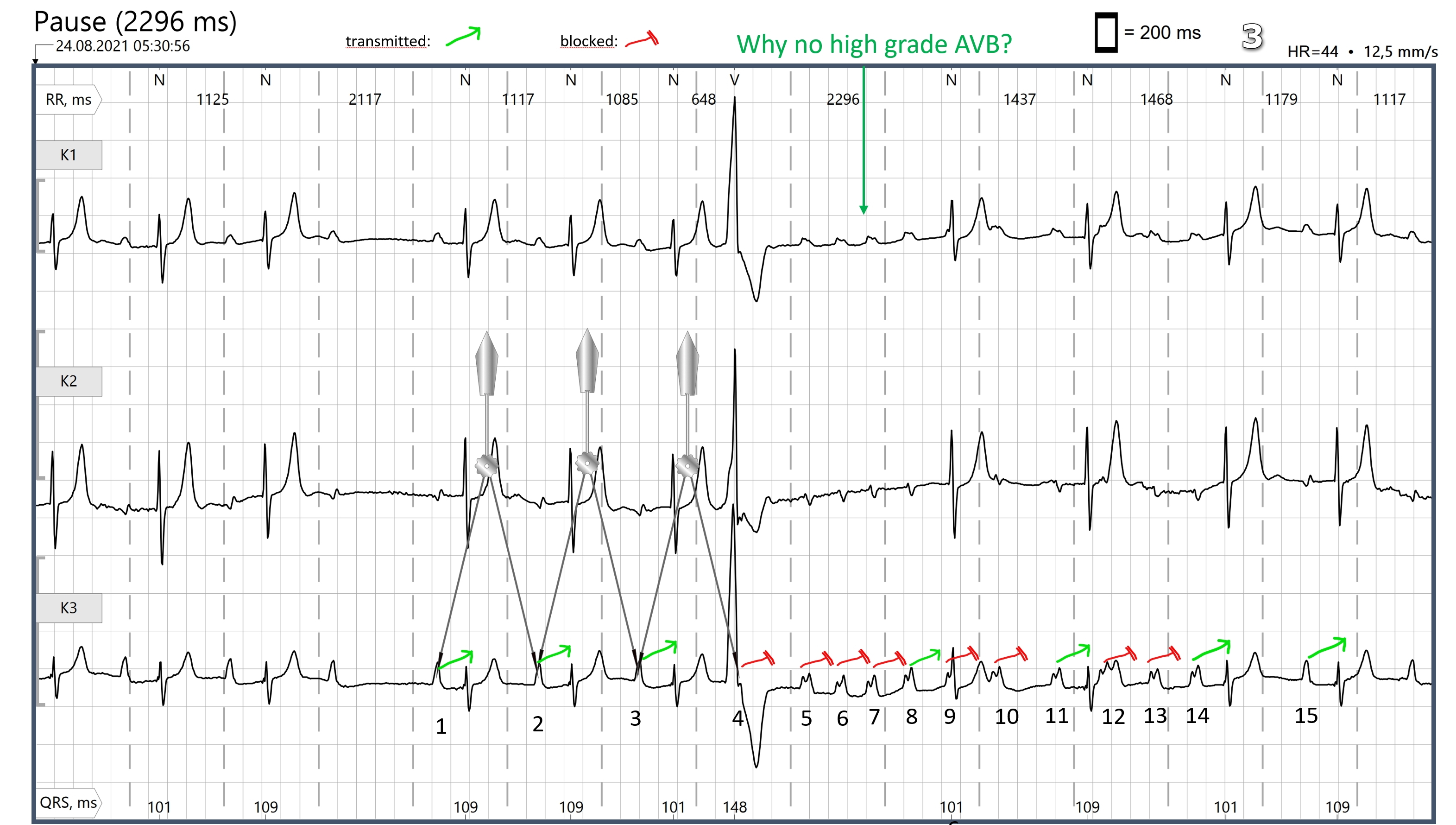
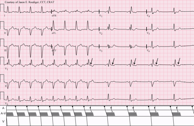
(Image 1) Why is there no second-degree AVB Mobitz type II and no high-grade AV block? To the first question: Basically, second-degree AV block Mobitz type II is rare. The two ECG patterns that can easily be confused with Type II Mobitz block are: blocked/non-conducted PACs and second-degree AVB Mobitz type I (Wenckebach). (Image 2) You have to compare the PR duration before the pause and after it. With the naked eye, the difference is often difficult to recognize, a pair of calipers does a good job here.
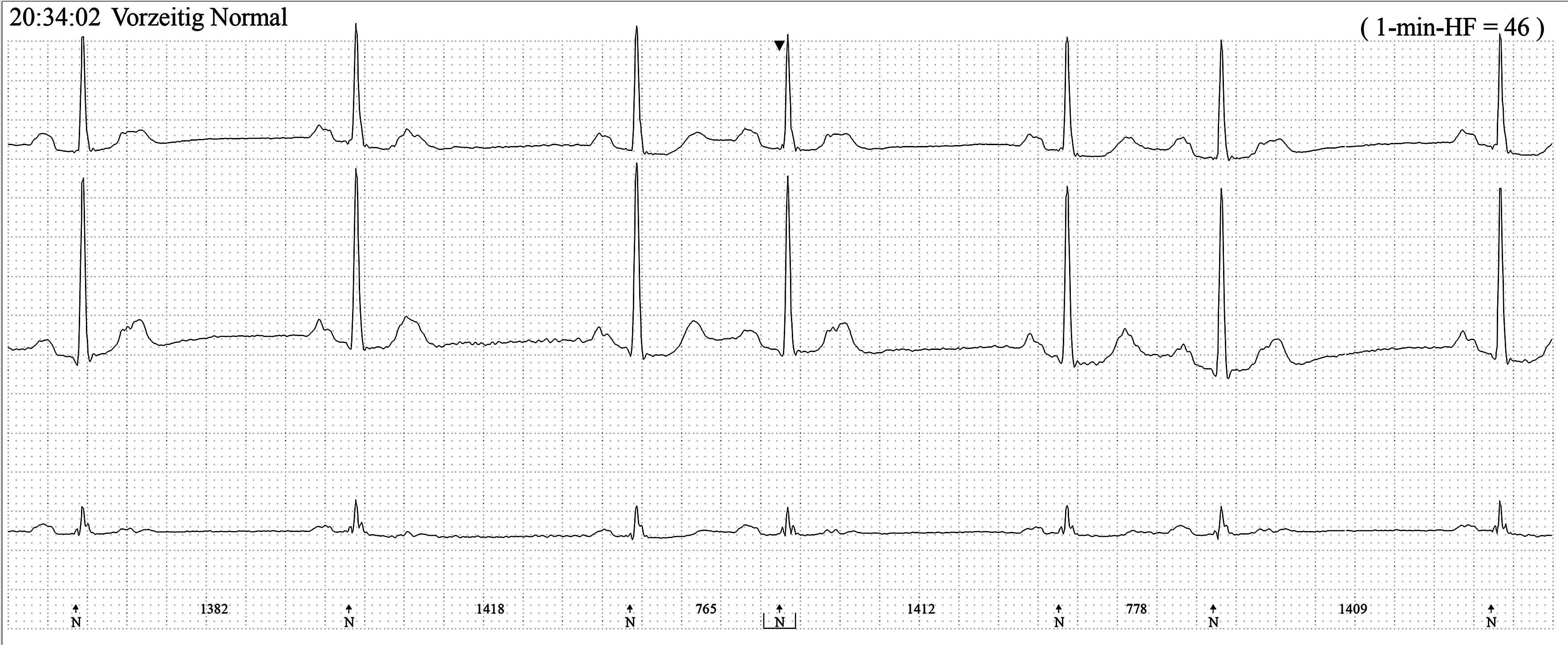
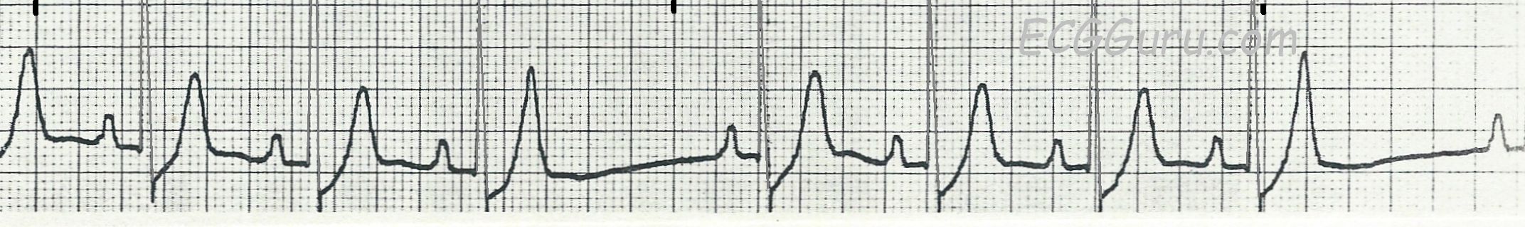
This is a good strip to demonstrate the change in the appearance of a T wave when a premature P wave occurs on the preceding T wave. The PACs found the atria ready to depolarize and produced a P wave that landed on top of the preceding T wave, making it appear taller than the others. The PACs also reset the sinus node, causing a slight delay before the next sinus discharge. The PACs occurred while the ventricles were still refractory, so no QRS complexes followed.