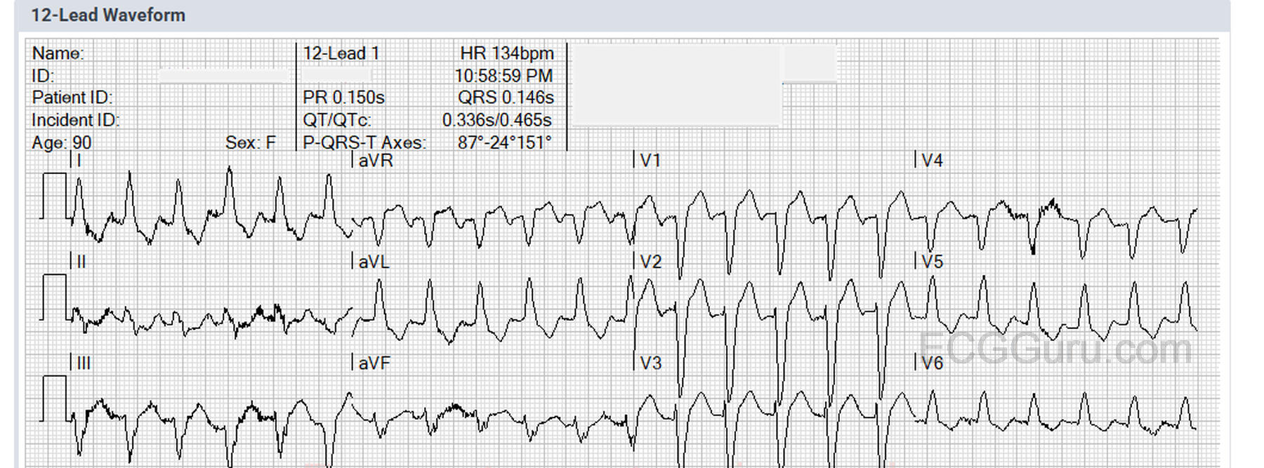
The Patient: This ECG was obtained from a 90-year-old woman who was complaining of feeling short of breath while lying in bed. Patient denied relevant medical history (respiratory or cardiac) and denied taking any medications. She also denied chest pain, nausea, or any other symptoms. She was found sitting upright in a recliner with mild subjective respiratory distress. She was completely alert and oriented, and was able to stand and transfer to the EMS stretcher. We do not have a record of her initial vital signs or lung sounds, but her initial SPO2 on room air was 80%. This improved to 100% on 15 L of O2 via NRB. She was transported to the hospital without incident.
The ECG: The ECG shows a rate of 134 bpm. The rhythm is regular. P waves are difficult to see because of the muscle tension artifact and the rate. I strongly suspect there is a P wave at the end of the T – best seen in aVR, in which you can see the negative P wave right after the T. The QRS is wide at 146 ms, and the QTc is borderline long at 465 ms, but is affected by the wide QRS being part of the measurement. There is, at this time, no simple and reliable method of evaluating QTc in the setting of wide QRS, but there are formulas available, and being evaluated.
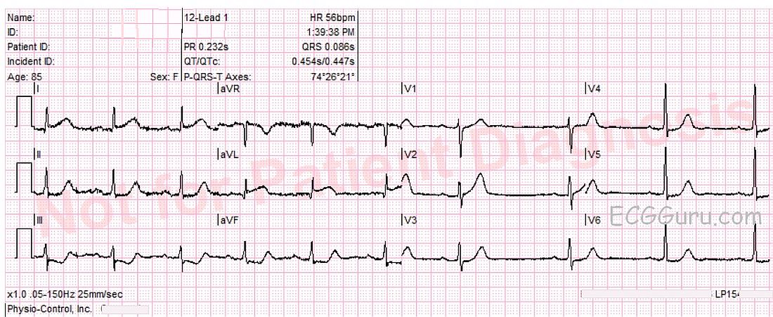
This ECG is taken from an elderly woman with chest pressure radiating to left shoulder for 30 minutes. She also complained of nausea with vomiting. Her family offered a history of unspecified cardiac disease, hypertension, hypercholesterolemia, and dementia.
The ECG shows ST elevation in Leads I and aVL, with reciprocal ST depression in Leads III and aVF. ST segments have an abnormally flat shape in Leads aVL, II, III, aVF, V5 and V6.
She was admitted to the hospital and sent to the cath lab. Her arteries were found to be clear, and Takotsubo cardiomyopathy was diagnosed. See also.
Our thanks to Lew Steinberg and Palm Beach Gardens Fire Rescue for donating this interesting ECG.
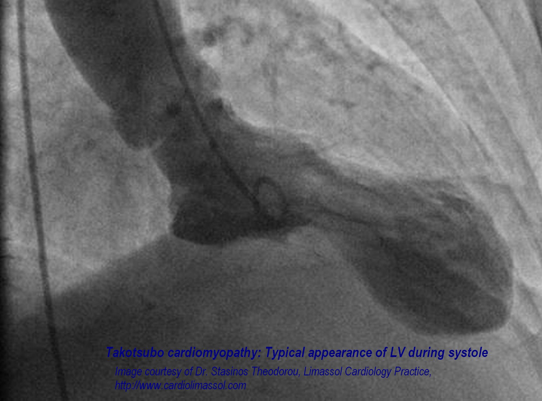
This image is courtesy of Dr. Stasinos Theodorou of the Limassol Cardiology practice. It may be used free of charge and free of copyright for educational purposes. For any commercial use, please contact Dr. Theodorou at the Limassol Cardiology practice.
This ventriculogram shows the typical apical ballooning of the left ventricle during Takotsubo myocardiopathy. This serious condition can be caused by extreme stress, such as in the death of a loved one, or prolonged high stress levels. The ECG will usually show ST elevations indicating an anterior wall M.I., but the angiogram will show no arterial occlusions. Takotsubo myopathy causes a sudden onset of congestive heart failure. It is transient, and those who survive it generally recover fully. For an excellent and complete discussion of Broken-Heart Syndrome,