Displaying 1 - 8 of 8
Dawn's picture

Narrow-complex Tachycardia In An Infant

The patient:  This ECG was obtained from a two-month-old girl who was a patient in the Emergency Department.  She had a fever due to a respiratory infection and was dehydrated. She was alert, active, and irritable.

The ECG:  There is a narrow-complex tachycardia at a rate of 194 bpm.  This is faster than the normal range for a two-month-old, which is about 80-160 bpm.  The intervals are all within normal range.  The frontal plane axis, at 145 degrees, is rightward, which is normal for this age. There are prominent, narrow Q waves in the inferior wall leads (II, III, and aVF) and in the left lateral leads (V4, V5, and V6).  There are no Q waves in the high lateral leads (I and aVL).  This is a normal pattern for this age group.   www.sciencedirect.com/science/article/pii/B9781416037743100280

The evaluation of this ECG must be preceded by a thorough evaluation of the patient.  SINUS TACHYCARDIA would be expected in the setting of fever, dehydration, hypoxia, pain or other discomfort. Should the rate fail to gradually return to a normal range after treatment, we would have to consider a reentrant supraventricular tachycardia. Reentrant tachycardias have a SUDDEN ONSET and SUDDEN TERMINATION.

Unfortunately, we do not have follow up on the patient.

Dawn's picture

Tachycardia In An Unresponsive Patient

 The Patient     This ECG was obtained from a 28-year-old woman who was found in her home, unresponsive.  She was hypotensive at 99/35.  No one was available to provide information about past medical history or the onset of this event.

Before you read my comments, pause to look at the ECG and see what YOU think.  We would welcome comments below from all our members!

The ECG     This ECG is quite challenging, as it illustrates the helpfulness of ECG changes in patient diagnosis, and also points out how important clinical correlation is when the ECG suggests multiple different problems. Forgive me in advance, but there is a lot to say about this ECG.

The heart rate is 148 bpm, and the rhythm is regular, although not perfectly. P waves are not seen, even though the ECG machine gives a P wave axis and PR interval measurement. The rate is fast enough to bury the P waves in the preceding T waves, especially if there is first-degree AV block. Differential dx: sinus tachycardia, PSVT, atrial flutter. The very slight irregularity points more towards sinus tachycardia.  The rate of nearly 150 suggests atrial flutter with 2:1 conduction, but the only lead that looks remotely like it has flutter waves is V2. The lack of an onset or offset of the rhythm makes it difficult to diagnose PSVT with any certainty.

jer5150's picture

Jason's Blog: ECG Challenge for the month of June, 2013.

Unfortunately, I have no available clinical data on this patient.  Merely looking for an interpretation of the ECG in it's raw form.

jer5150's picture

Jason's Blog: ECG Challenge of the Week for Sept. 9-16.

Patient's clinical data:  76-year-old white man admitted to the ICU.  

Hint:  In Fig. 2, there is an extremely subtle clue on that ECG that I almost didn't notice.  Laddergrams will be provided for both of these as the end of the week.

What is going on here?  


jer5150's picture

All depends on how you define "degrees".

One of our local news channels was reporting widespread and record-breaking right axis deviation throughout central Virginia.  

References / Sources:
1.)  WRIC Richmond News and Weather - - WRICTV8 - Home
2.)  Blaufuss Multimedia - Heart Sounds and Cardiac Arrhythmias

jer5150's picture

Jason's Blog: ECG Challenge of the Week for June 17-24. What "highs" and "lows" is this tracing suggestive of?

This week’s ECG is categorized under the heading:“Tracing suggestive of          This is a classical example of this pair of derangements.

Dawn's picture

Question: Does an extreme right axis (backward) always indicate a ventricular rhythm?

Today's expert is Jason E. Roediger, CCT, CRAT, who is a highly respected Cardiovascular Technician at the Dept. of Veterans Affairs, Hunter Holmes McGuire VA Medical Center in Richmond, VA. He is known for holding numerous certifications in all levels of ECG interpretation, and also for scoring 100% on the Level IV Advanced ECG Board Certification exam that is usually reserved for cardiologists.
 

Answer: Always? No. Usually, Yes.
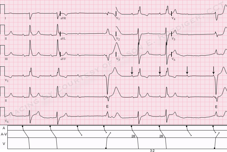
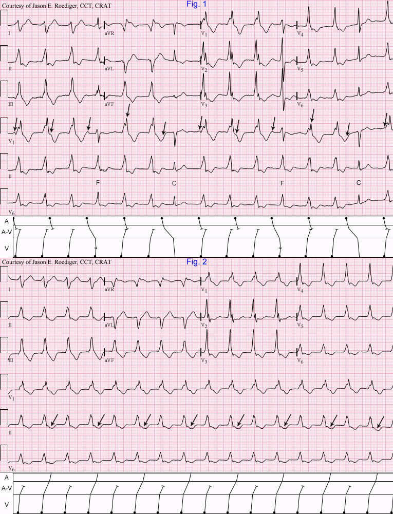
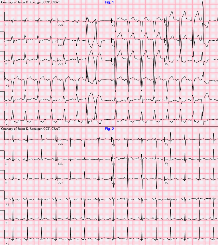
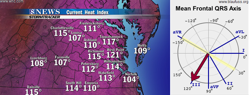
There are exceptions to several "golden rules" in electrocardiography and this one is not exempt. One of the chronic issues contributing towards widespread confusion in understanding electrical axis is a lack of continuity in terminology. There is no general concensus on how to refer to an axis in the right upper quadrant. Depending on which author you are reading, it has traditionally been known by multiple names: Northwest axis. . . upper right quadrant. . . extreme right axis. . . right superior axis. . . "no-man's-land" (i.e., "N-M-L".). . . etc. Because my first exposure to electrical axis was through Dr. Marriott's textbooks and he prefered to use "N-M-L", I have personally latched on to that particular name as well. Even though some persist in calling it an "extreme left axis" or "far left axis deviation", this practice is frowned upon and discouraged. It's important to note that an axis in "N-M-L" is not synonymous with an "indeterminate" axis which occurs when the QRS is essentially isodiphasic or equphasic in all 6 limb leads and therefore the polarity of the QRS cannot be discerned in leads I and aVF.

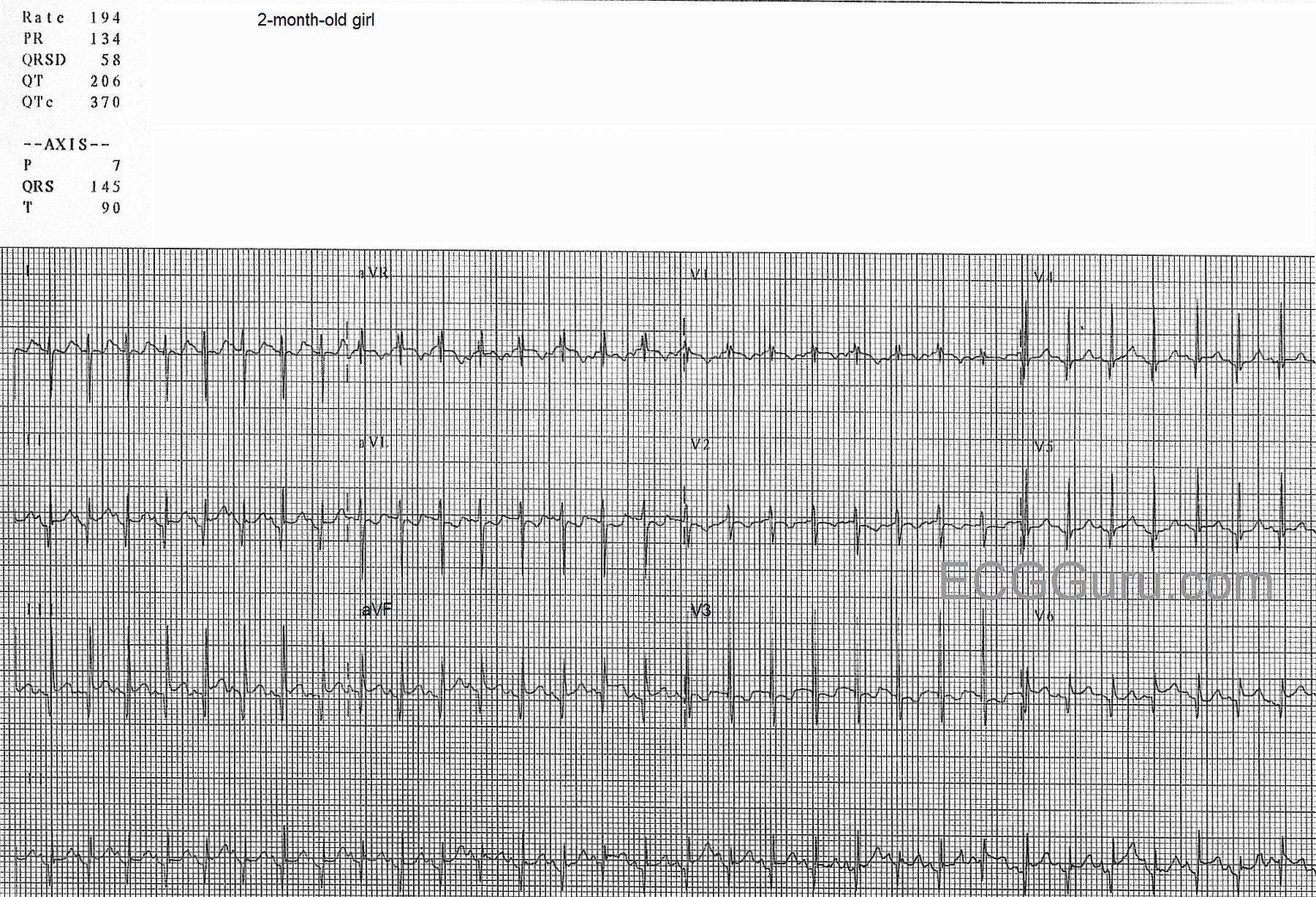
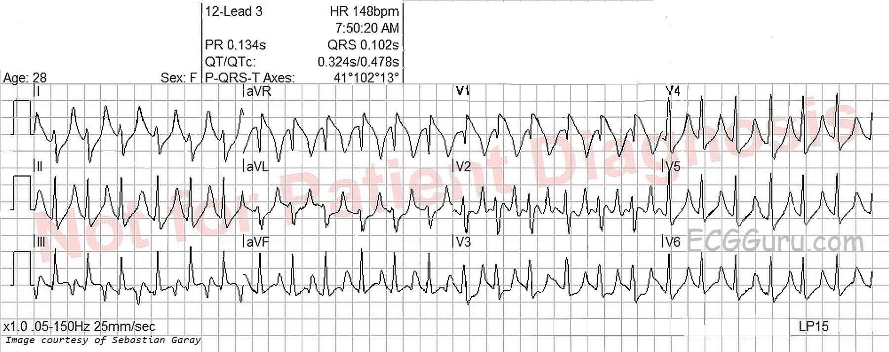
Definition: An axis in "N-M-L" is recognized when the QRS complex has a predominantly or wholly negative deflection (i.e., down) in leads I and aVF. The axis is −90 to −180 degrees.

Irregardless of which descriptive name you prefer, in the context of a wide QRS complex tachycardia, this particular axis is highly predictive of ventricular tachycardia and is rarely encountered in "conducted" rhythms however some examples of aberrant SVT have been published with an axis in "N-M-L".

In summary: An axis in "N-M-L" implies (but is not proof of) an apical origin to the rhythm and should make one think of and exclude the possibility of ventricular tachycardia. As a general rule, until it is proven otherwise, assume any wide QRS complex tachycardia is ventricular tachycardia. Even though this one clue carries significant weight in supporting the interpretation of ventricular tachycardia, that conclusion can not be made based solely on this single criteria. This axis is just one of a long list of criteria and should be used in conjunction with all of them as they carry alot of strength when used collectively.
 

All our content is FREE & COPYRIGHT FREE for non-commercial use

Please be courteous and leave any watermark or author attribution on content you reproduce.