Displaying 1 - 3 of 3
Dawn's picture

ECG Basics: Paroxysmal Supraventricular Tachycardia Treated With Adenosine

This series of ECG rhythm strips shows a paroxysmal supraventricular tachycardia successfully treated with adenosine.  The patient was complaining of a rapid heart rate and palpitations, but was hemodynamically stable.  It is not known whether any parasympathetic stimulation, such as a Valsalva maneuver or carotid sinus massage, was used initially.   

The first rhythm strip shows a PSVT, presumably AV nodal reentrant tachycardia, at a rate of about 215 per minute.  (We originally indicated a rate of 240 per minute, but this was a typo). Using the simplist method of determining rate, the six-second method, we see 21, but almost 22, QRS complexes in six seconds. Differential diagnosis would include sinus tachycardia, but this rate is too fast for sinus tach, especially in a resting patient.  Also, sinus tach would slow down as the patient is rested or made more comfortable, and this rate did not vary.  Also, when confronted with a supraventricular tachycardia, one should also consider atrial flutter and atrial fibrillation.  This is somewhat slow for atrial flutter with 1:1 conduction, and that rhythm is much more rare than AVNRT. It is too regular for atrial fibrillation.  So, we are left with the probable diagnosis of paroxysmal supraventricular tachycardia.  The “paroxysmal” part is presumed since AVNRT has an abrupt onset, and the patient’s symptoms started suddenly. 

The second rhythm strip shows what happened after adenosine was administered.  The patient received first a 6 mg dose, rapid IV push.  When that was not effective, he received 12 mg rapid IV push.  The rhythm strip is typical of the first minute or so after adenosine administration.  Adenosine can cause transient AV blocks, escape rhythms, and ectopic irritability.  The half-life of adenosine is only 6 seconds, so the dysrhythmias and uncomfortable symptoms are short-lived.  In this strip, we see frequent PVCs and runs of V tach.  

Dawn's picture

ECG Basics: Paroxysmal Supraventricular Tachycardia Converted With Adenosine

Today, you get THREE strips for your basic classes!  The first shows a PSVT - paroxysmal supraventricular tachycardia at a rate of about 220/minute.  The QRS complexes are narrow, and the rhythm is regular.  In the second strip, we see the moment of conversion after a dose of 6 mg. of adenosine was administered by rapid I.V. infusion.  The re-entry cycle is broken, and the patient experiences many PVCs, including groups of two, three, and even four in a row.  Soon, however, the rhythm settles into sinus rhythm with PACs, and later, just sinus rhythm (not shown).  For your more advanced students, the second strip shows the PVCs interacting with the underlying sinus rhythm.  There are several fusion beats with varying degrees of fusion, and one can sometimes see a sinus P wave just before a PVC.  None of this is clinically significant in this patient, because the ventricular ectopy was a side effect of the treatment, and was self-limited.  It is a great strip for teaching, though! 

Dawn's picture

Wide-Complex Tachycardia Converted by Adenosine

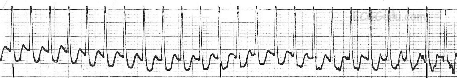
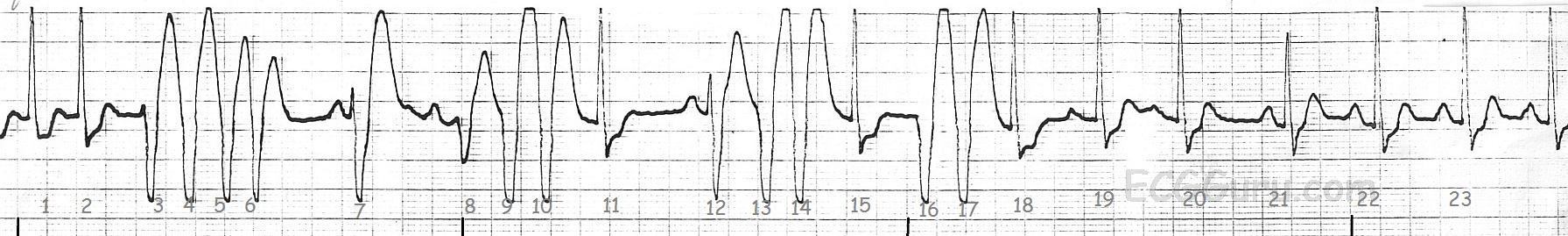
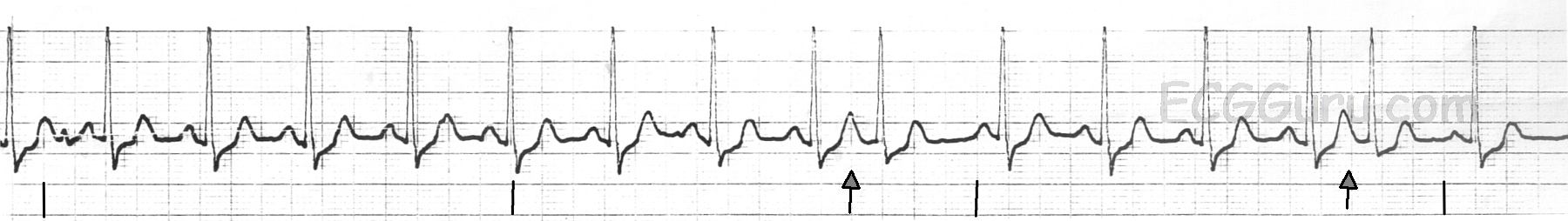
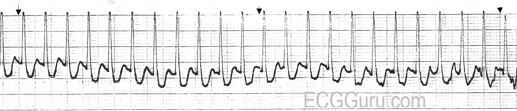
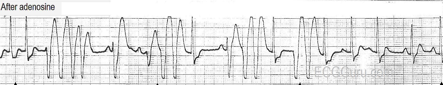
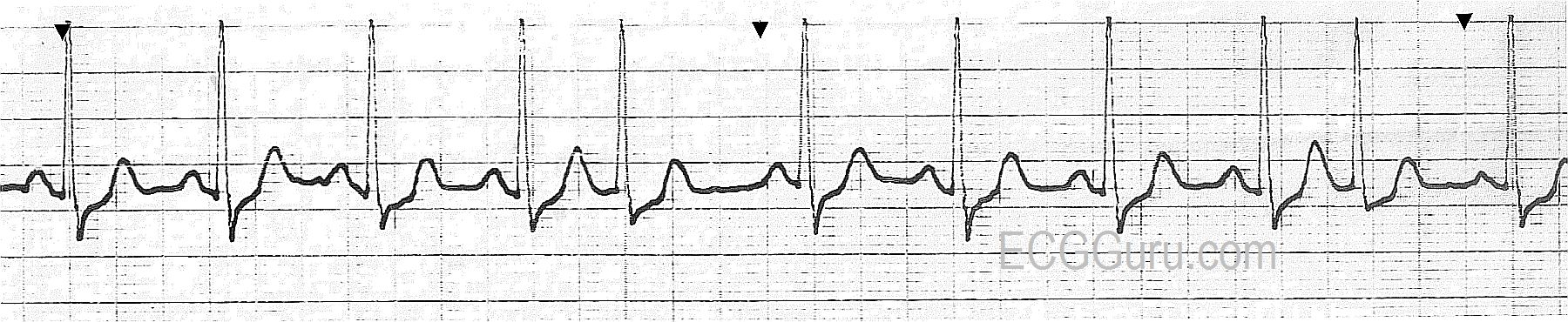
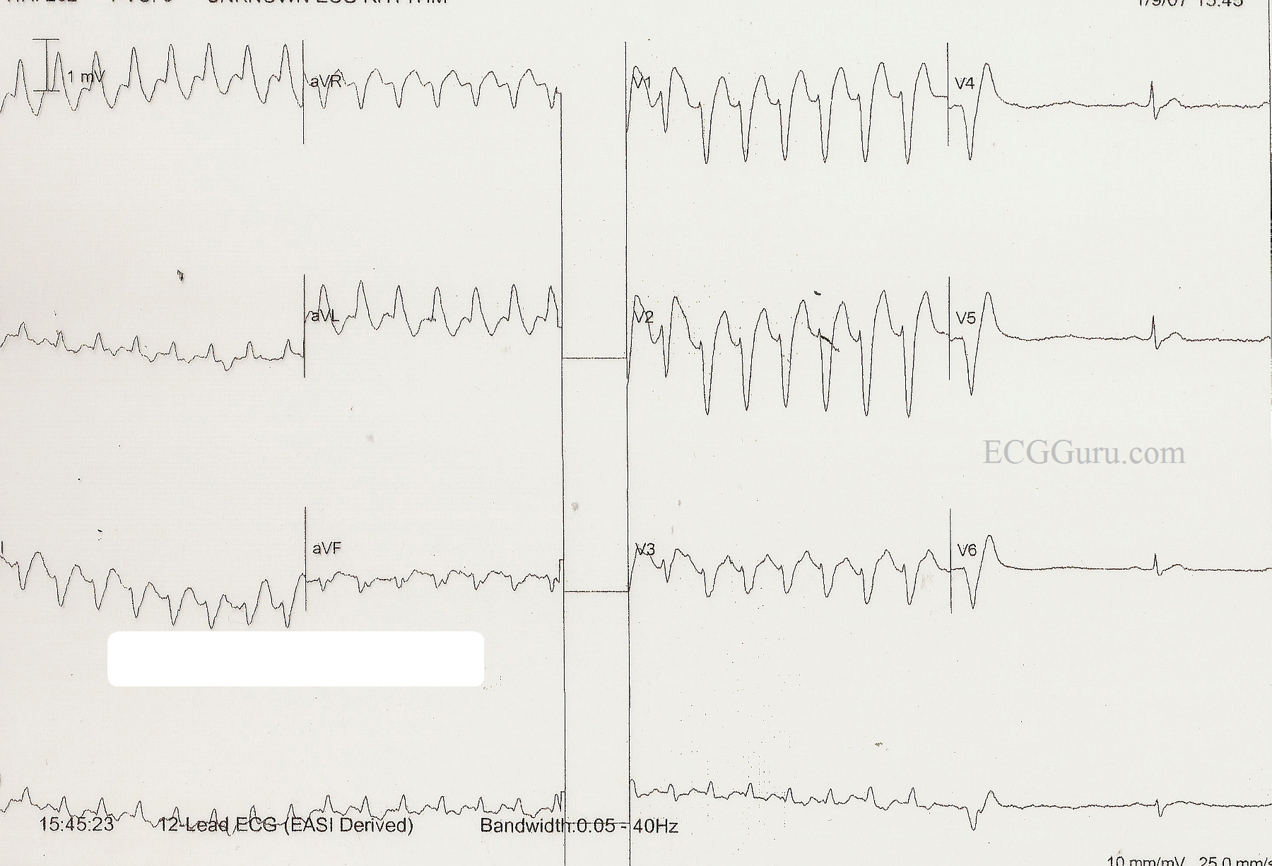
This ECG was presented earlier this week as an example of SVT with LBBB aberrancy, which was ultimately converted with one dose of adenosine in the Emergency Department.  It is the most shared and commented on ECG yet to appear on the Guru.  The diagnosis given was the one accepted by the medical staff who cared for the patient, who was a man in his 30's who presented to the Emergency Department complaining of a rapid heart rate.  He was ambulatory with stable vital signs, in spite of the tachycardia. He reported that he has had several episodes of fast heart rate which responded to either Valsalva maneuvers or, in some cases, medication in the ED.  He was told he might benefit from an ablation procedure, but he did not have health insurance and continued to use the ED as his primary source of medical care.  When he was admitted to the ED, the tech initially called for help, thinking the monitor showed ventricular tachycardia.  The ED physician felt that this represented LBBB aberrancy, possibly rate-dependent, and he treated the patient with adenosine.  The rhythm converted to sinus after one dose, and the patient remained stable throughout the process.  He was advised to undergo further observation and testing, but he declined due to financial concerns, and the fact that he usually succeeded in relieving his symptoms with "bearing down".  

Wide-complex tachycardias can be difficult to assess simply from an ECG. The patient's stability depends more upon general health and cardiac output issues than the origin of the tachycardia.  When we presented this ECG, we also presented the diagnosis he had upon discharge from the ED.

Subsequently, ECG Guru Dr. Ken Grauer, a frequent contributer to this site, offered his alternative diagnosis and his explanation of why he believes this to be v tach.  Other well-respected ECG experts have also questioned the original diagnosis.  Please refer to the comments below for this very helpful explanation.  Unfortunately, this patient is lost to followup, as this incident occurred some time ago.

WTCs remain a most fascinating topic, especially for those who enjoy "detective work".  We thank Dr. Grauer, Tom Bouthillet, and others for their contributions to the ECG Guru on this topic.
   

All our content is FREE & COPYRIGHT FREE for non-commercial use

Please be courteous and leave any watermark or author attribution on content you reproduce.