Displaying 1 - 4 of 4
Dawn's picture

Bigeminal Rhythm With Aberrant Conduction

This ECG is a good example of sinus rhythm with aberrantly-conducted PACs.  The tracing was donated to the ECG Guru several years ago by Dr. Ahmed from Sanjiban Hospital in India.  We have no patient data for this tracing. 

The underlying rhythm here is normal sinus rhythm. Most of the parameters – rate, PR interval, and QRS duration – are normal.  The QTc interval, which is the QT interval corrected to a rate of 60 bpm, is prolonged at 568 ms.  We do not know the patient’s clinical condition or medications, so we cannot guess at the reason.  However, a prolonged QTc is associated with an increased risk of Torsades de pointes, a type of polymorphic ventricular tachycardia. 

The first three beats appear the same (Leads I, II, and III).  However, the first R-to-R interval is shorter than the second one.  This could be due to rate variation, a concealed sinus block, or a premature atrial contraction (PAC).   The P wave of the “early” beat, marked #1, looks slightly different from the other P waves in Lead II, but, because of the slow rate, there is no way to be sure without a longer rhythm strip.  After the possible PAC, the rhythm becomes coupled, probably atrial bigeminy, where every other beat is a PAC.  There are several mechanisms that cause grouped beating, but atrial ectopic bigeminy is the most common. Normally, PACs have different-looking P waves compared to the sinus beats.  In this ECG, the P waves are often buried in the preceding T waves, and are hard to evaluate. 

Dawn's picture

Atrial Bigeminy Vs Possible Sino-atrial Exit Block

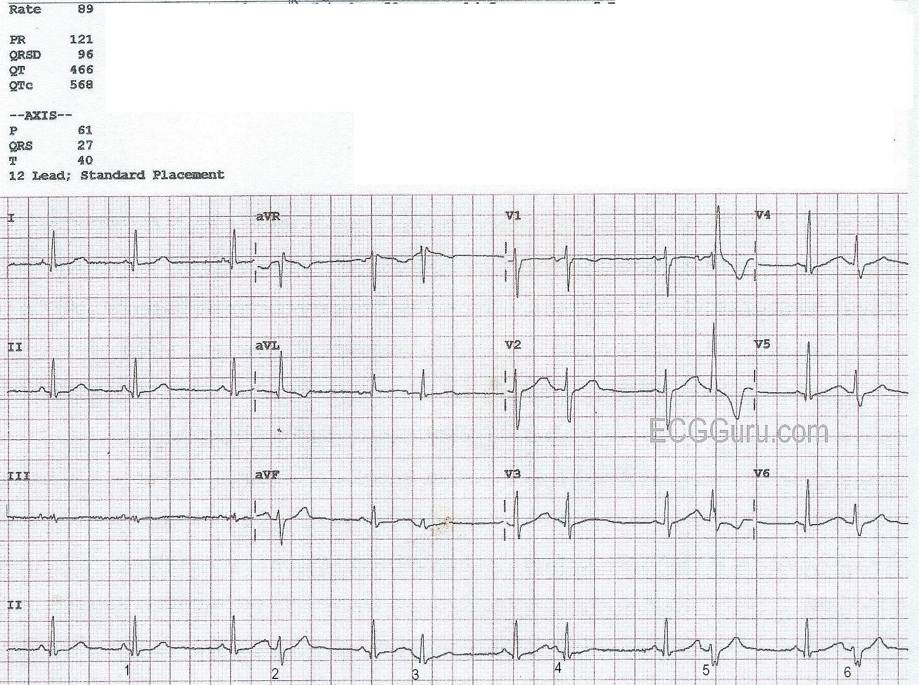
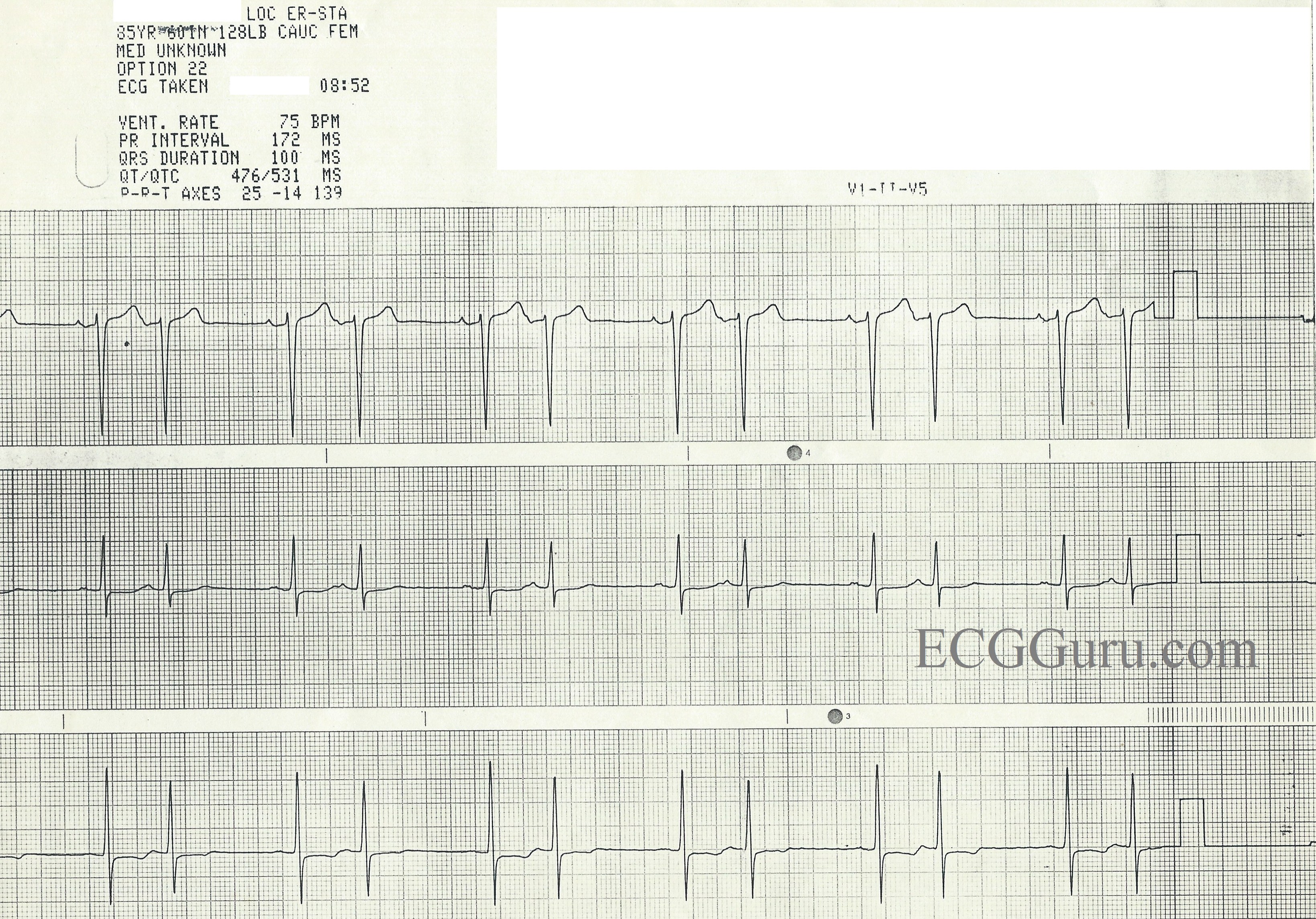
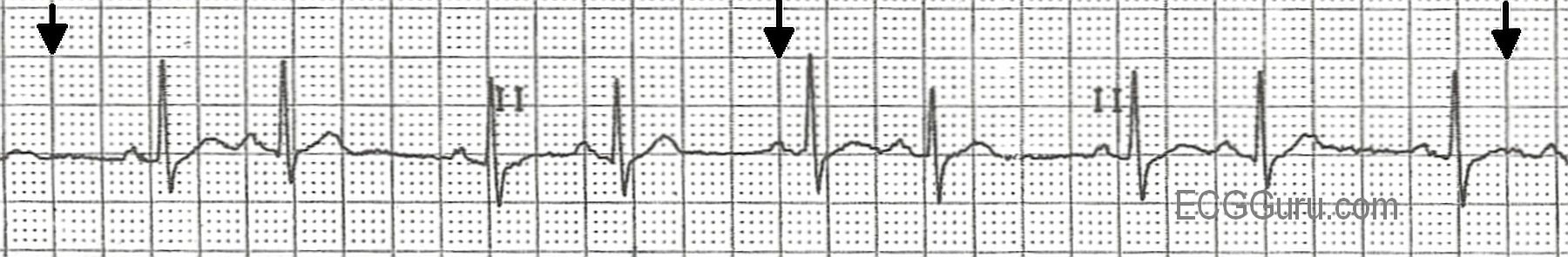
This three-lead rhythm strip is from an 85 year-old-woman for whom we have no other information, unfortunately.  It shows an obvious, regular bigeminal pattern.  There are many rhythms that can cause bigeminy, with regular, paired beats.  So, how do we analyze this rhythm? 

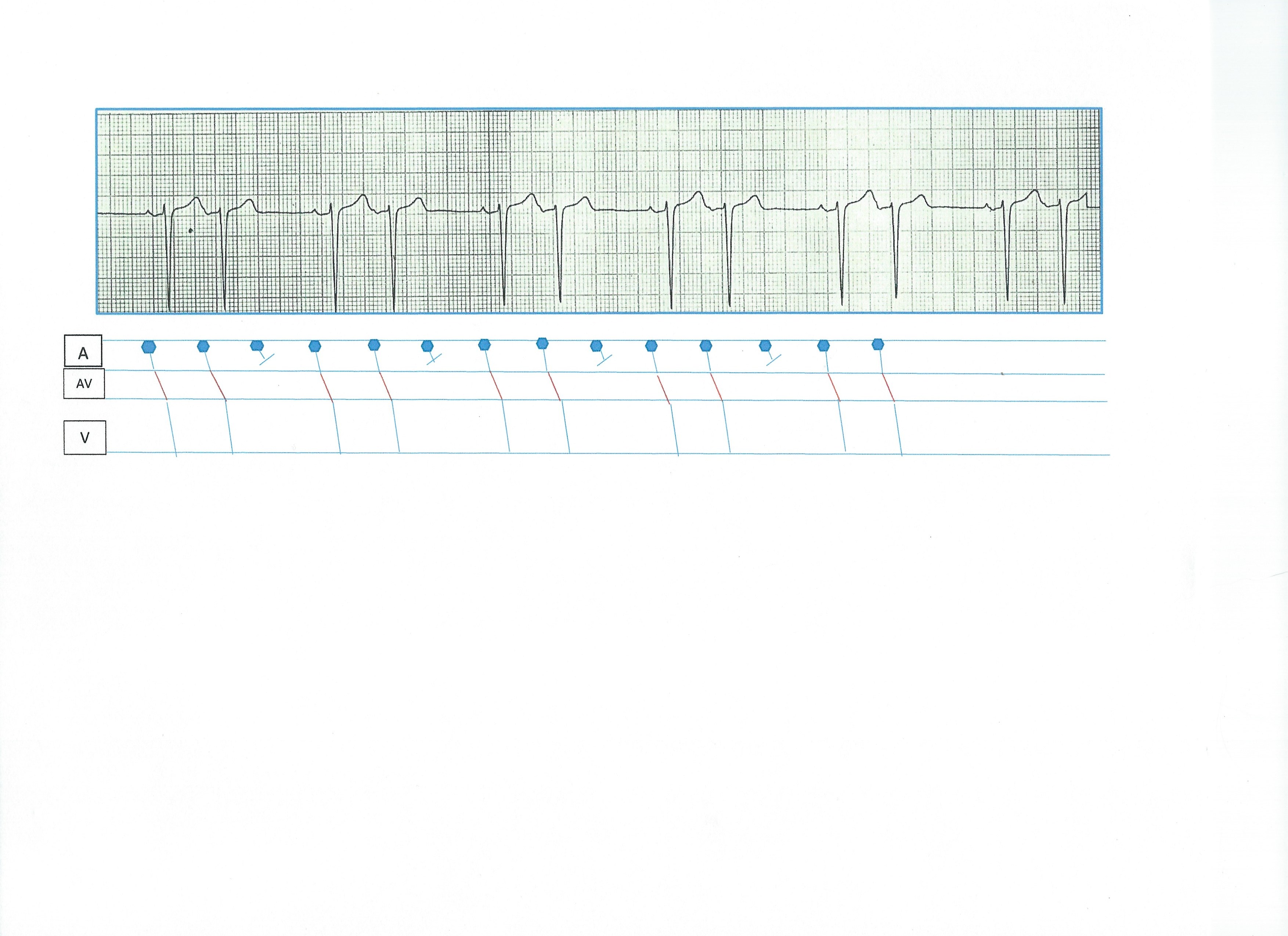
First, we can see that all the QRS complexes are fairly narrow and look alike in each lead.  So, we know this is a supraventricular rhythm.  The rhythm “marches out”, with the same spacing between all the paired beats, and exactly one P-QRS missing after each pair. 

Sino-atrial exit block occurs when the sinus node fires, but the impulse is unable to exit the sinus node. No P wave will occur, but the sinus node continues to fire in a regular rhythm.  There are four types of SA block, just as there are four types of AV block.  This ECG meets the criteria for a Second-degree, Type II SA block. 

First-degree SA block would not be decipherable on the ECG, as we do not see the SA node fire, and therefore cannot measure an interval between the SA node firing and the production of a P wave. 

Second-degree SA block, Type I would have progressively prolonging exit times for the impulse leaving the SA node.  This would produce progressively shortening R-to-R intervals, until the pause. 

Second-degree SA block, Type II has regular R-to-R intervals until the pause, and the pause will equal two or more (usually two) R-to-R intervals. 

Third-degree SA block produces NO P WAVES, and would have to be diagnosed on electrophysiology studies, as it would usually result in a junctional escape rhythm. 

Dawn's picture

Sinus Rhythm With Atrial Bigeminy

This ECG is from an 88-year-old man with congestive heart failure.  No other clinical information is known.  It shows an underlying sinus rhythm with atrial bigeminy - every other beat is a premature atrial contraction.  There is very little, if any, difference in the morphology of the sinus P waves and the ectopic P waves, indicating that the ectopic focus is in the vicinity of the sinus node.  There is no "compensatory" pause, because PACs penetrate the sinus node, resetting it.  So, the underlying sinus rate here is about 72 beats per minute.  There are several mechanisms for bigeminy to occur, but ectopic bigeminy is the most common.

Dawn's picture

ECG Basics: Sinus Rhythm With Atrial Bigeminy

This is a normal sinus rhythm with atrial bigeminy, a term meaning that every other beat is a PAC.  If you look carefully, you can see slight differences in the sinus P waves and the atrial (premature) P waves.  The PACs penetrate and reset the sinus node, causing what looks like a delay after the PAC.  It is often just a return to the normal P to P interval, or nearly so.  If you teach basic students in a clinical setting, they will learn from palpating the peripheral pulse and feeling the pattern of bigeminal beats. Sometimes, the premature beat feels much weaker due to less filling time available to the ventricles.  Atrial bigeminy can have very benign causes, such as increased caffeine intake, or it can have more complex causes such as advanced heart disease or conduction blocks.  In some patients, atrial bigeminy, or any PACs, can be a precursor to more serious atrial dysrhythmias, such as atrial fibrillation.

All our content is FREE & COPYRIGHT FREE for non-commercial use

Please be courteous and leave any watermark or author attribution on content you reproduce.