The Patient:This ECG was obtained from a man in his mid-sixties who was complaining of chest pain. The pain had an acute onset and is described as "10" on a 1-10 scale. He has a PMHx of coronary artery disease with stents in his right coronary artery and minimally invasive aortic valve replacement.
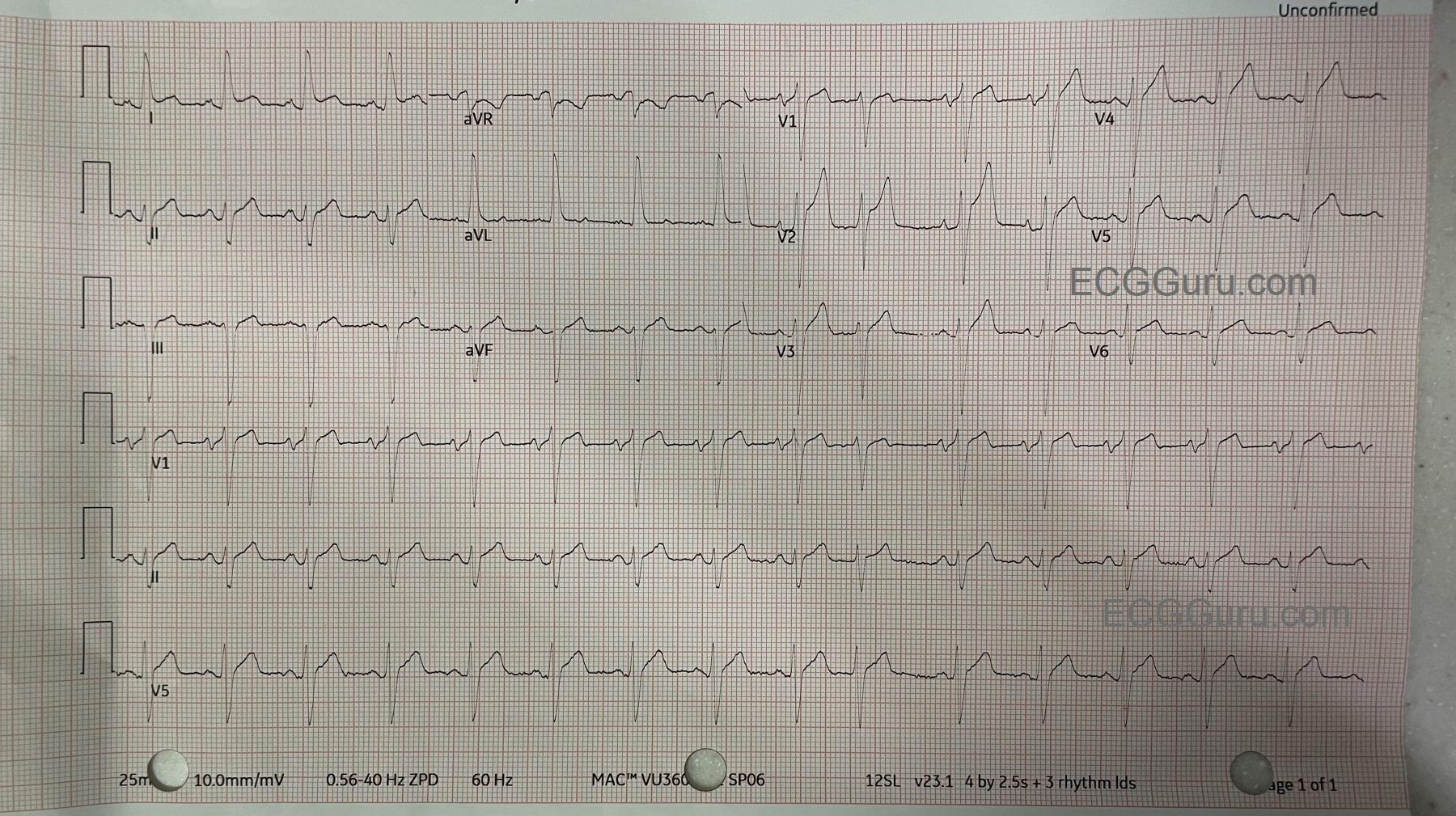
The ECG: The rate is 86 bpm. The rhythm is normal sinus rhythm with one PAC (10th beat). The PR interval is .18 seconds (176 ms), the QRS duration is .122 seconds (.12 seconds). This represents a ventricular conduction delay. There is no right or left bundle branch block. The QT/QTc is 333 ms/400 ms (B). The frontal plane QRS axis is leftward, with criteria for left anterior fascicular block. LAFB can be explained by this patient's history of prior CAD and valve replacement. There is ST elevation in Lead I and also in V1-V6. The ST segments have a straight shape in Leads I and aVL and in V1-V6. This shape represents ischemia in a patient with these symptoms and ECG findings. This is an ANTERIOR-LATERAL OCCLUSIVE M.I.
Followup: The patient was taken to the cath lab and had angioplasty of an occlusive mid-LAD (left anterior descending) lesion and a partially-occlusive mid-RCA lesion.