Displaying 1 - 3 of 3
Dawn's picture

Large Anterior Wall M.I. and Effect of Lead Reversal

EDIT: Please refer to the comments below this text. The second ECG in this series shows unexpected QRS and ST-T morphology changes, which I tried to explain by way of the patient's long anterior descending coronary artery. However, Dave Richley, who is a very well-known cardiac physiologist and ECG Guru took the time to analyze these morphologies and realize they can be explained by an inadvertent ECG LEAD MISPLACEMENT. This patient does have a proximal lesion of the LAD, proven and repaired in the cath lab. But the inferior wall does not have the injury it appears to have in this second ECG. Thanks to Dave for reminding us to slow down and look closely when things don't look "right".

The Patient:   These two ECGs, taken 26 minutes apart, were obtained from a 50-year-old man who complained of sudden onset of chest pain.  He denied history of coronary artery disease. He was Covid-positive, and the rest of his medical history was unremarkable.

ECG No. 1:  This ECG was obtained by paramedics enroute to the hospital.  For your beginner-level students, it will be easy to demonstrate the large ST elevations in V3 through V6. The machine’s measurements at the bottom confirm that this ECG meets any field criteria for ST elevation M.I. “STEMI”.

But there is so much more to see! Taking a methodical approach, and starting with rate and rhythm, we see sinus bradycardia at 57 bpm. Intervals and frontal plane axis are within normal limits. R wave progression in the chest leads is stalled in V1- V3 due to loss of initial r waves (narrow QS). The transition to positive deflections in V4 – V6 is abrupt.  These q waves in the V1 and V2 appear narrow, but V3 appears to have a Q wave that is almost wide enough to be considered pathological.  Narrow Q waves may be a transient sign of injury, while wide ones (>40 ms) are an ECG sign of necrosis.

Dawn's picture

Lateral Wall M.I.

The patient:   This ECG was taken from a 66-year-old man who was complaining of chest pain at rest. He had been previously diagnosed with lung cancer with metastases to his bones.  The last ECG, taken one week ago, was normal.

The ECG:  There is mild sinus tachycardia at 101 bpm.  The rhythm is regular.  The QRS duration and PR interval are normal, as is the QTc.  The QRS voltage in the limb leads is small, and we do not know the patient’s height and weight.

There are notable ST elevations in I and aVL (high lateral wall) and in V5 and V6 (low lateral wall).  When the high and low lateral walls are similarly affected, we usually look to the circumflex artery as the culprit artery.  We also see ST depression in Leads III and aVF (reciprocal to the STE in I and aVL) and in V1 – V4.  This could indicate subendocardial damage or reciprocal changes.  This ECG meets the criteria for acute lateral myocardial infarction.

The patient was taken to the cath lab emergently.  His coronary arteries, including the left circumflex, all were free of occlusive lesions.  He had no coronary spasm during the procedure, but it was decided that spasm had been the cause of the ECG changes.  His ECG reverted to normal.

It is important to record abnormal findings, as some changes can be temporary or fleeting.  Coronary artery spasm can cause ischemia and damage to the heart, just as plaque lesions and blood clots can.

Dawn's picture

Circumflex Occlusion with Posterior-lateral M.I.

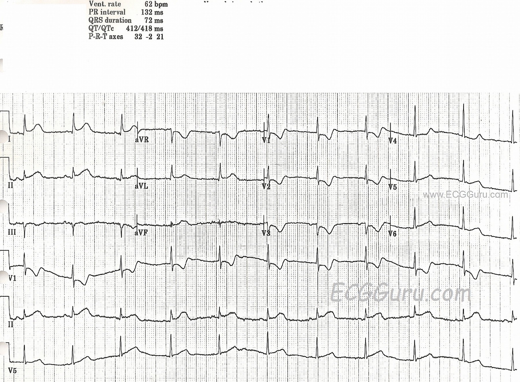
This ECG was obtained from a woman with chest pain who was taken to the cath lab and found to have a 100% occlusion of her circumflex artery.  

There are obvious ST segment elevations in Leads I and aVL, as well as in Lead II.  Lead II is the most leftward of the inferior wall leads, and I and aVL reflect the high lateral wall. She also has ST depressions in V1 through V3.  If you look closely at the R wave progression in the anterior leads, you will readily note that it appears that V1 and V3 wires have been reversed.  That being said, the "real" V2 and V3 have taller-than-normal R waves.  The tall R waves and ST depression are signs of "posterior wall M.I."  Recently, the actual definitions of the "lateral" wall and "posterior" wall have come into question.  However, the important thing clinically, is that this patient IS experiencing an ST elevation M.I. (STEMI), which was confirmed in the cath lab.  The locations of the ST changes were consistent with the 100% occlusion of her circumflex artery.

For our more advanced readers (and our "Gurus"), there is an interesting rhythm.  The P wave morphology changes frequently, even though the rhythm remains regular.  The rate, at 62 BPM, was adequate, and the patient did not suffer any consequential dysrhythmias during her procedure.  We don't have long-term followup information on her.


All our content is FREE & COPYRIGHT FREE for non-commercial use

Please be courteous and leave any watermark or author attribution on content you reproduce.