Displaying 1 - 3 of 3
Dawn's picture

Acute Occlusive Myocardial Infarction With Cardiogenic Shock

The Patient:  67-year-old man complaining of chest pain radiating to his jaw, 10/10. He is short of breath and diaphoretic. We do not know his BP, just that it was low.  The patient states “no past medical history – never hospitalized”. He thought himself to be very healthy.  He was given aspirin 325 mg and transported to a full-service cardiac hospital as a “cardiac alert”.

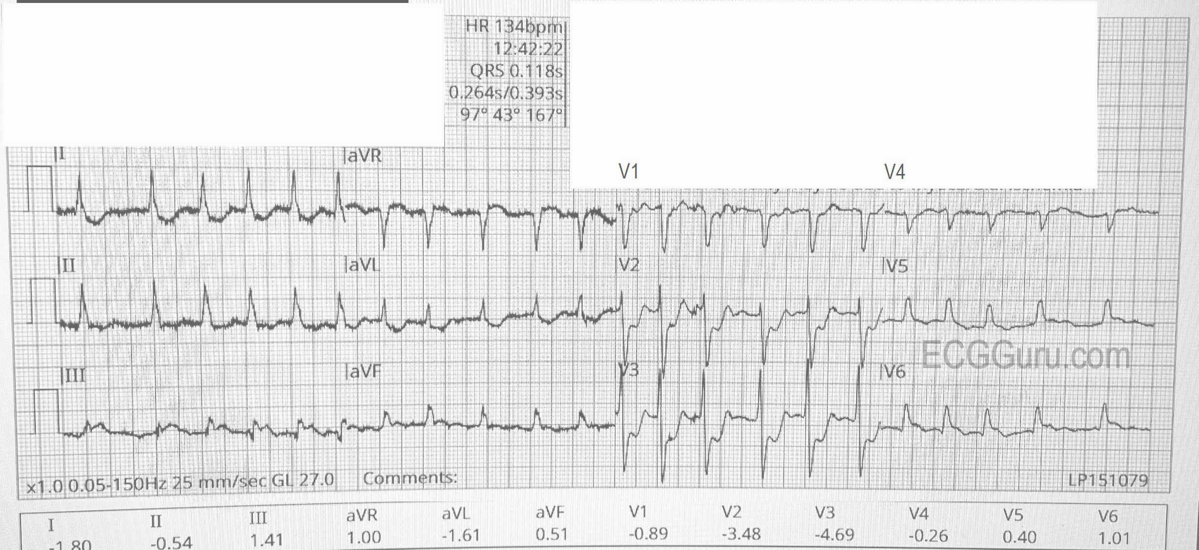
The ECG (from EMS): 

The rhythm is atrial fibrillation with a rapid ventricular response (about 134/min.). The QRS width is .118 seconds (118 ms).  The frontal plane axis is slightly to the left, but WNL. The R wave progression mostly normal, but V4 is incongruous.  V2 and V3 have a tall R wave, possibly representing a pathological Q on the posterior side.  There are ST CHANGES in every lead. ST elevation is noted in III, aVF, aVR, V5 and V6, representing ischemia in the inferior wall. There is ST depression in all other leads, indicating widespread subendocardial ischemia and/or acute reciprocal depression.  Interesting that Lead II would normally be elevated when III and aVF are, but aVR is elevated, causing reciprocal ST depression in Lead II.  So, Lead II looks almost normal.  

The pattern of ST elevation in aVR with widespread ST depression can indicate:

1)     Proximal occlusion of LAD or significant stenosis of Left Main artery.

Dawn's picture

Anterior-lateral M.I.

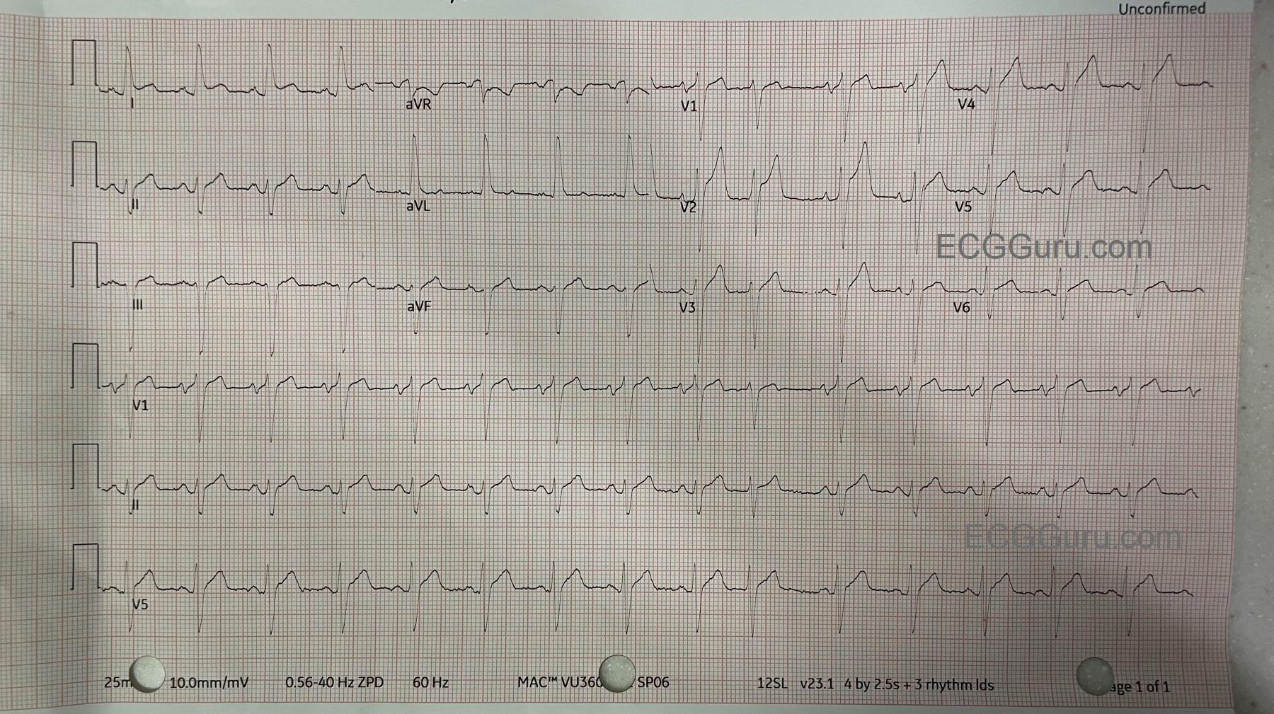
The Patient: This ECG was obtained from a man in his mid-sixties who was complaining of chest pain.  The pain had an acute onset and is described as "10" on a 1-10 scale.  He has a PMHx of coronary artery disease with stents in his right coronary artery and minimally invasive aortic valve replacement.

The ECG:  The rate is 86 bpm.  The rhythm is normal sinus rhythm with one PAC (10th beat).  The PR interval is .18 seconds (176 ms), the QRS duration is .122 seconds (.12 seconds). This represents a ventricular conduction delay. There is no right or left bundle branch block.   The QT/QTc is 333 ms/400 ms (B). The frontal plane QRS axis is leftward, with criteria for left anterior fascicular block. LAFB can be explained by this patient's history of prior CAD and valve replacement.  There is ST elevation in Lead I and also in V1-V6.  The ST segments have a straight shape in Leads I and aVL and in V1-V6.  This shape represents ischemia in a patient with these symptoms and ECG findings.  This is an ANTERIOR-LATERAL OCCLUSIVE M.I.

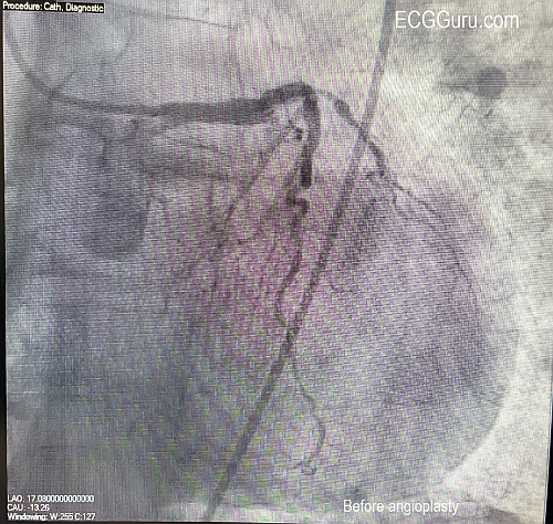
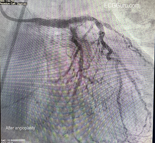
Followup:   The patient was taken to the cath lab and had angioplasty of an occlusive mid-LAD (left anterior descending) lesion and a partially-occlusive mid-RCA lesion.  

Dawn's picture

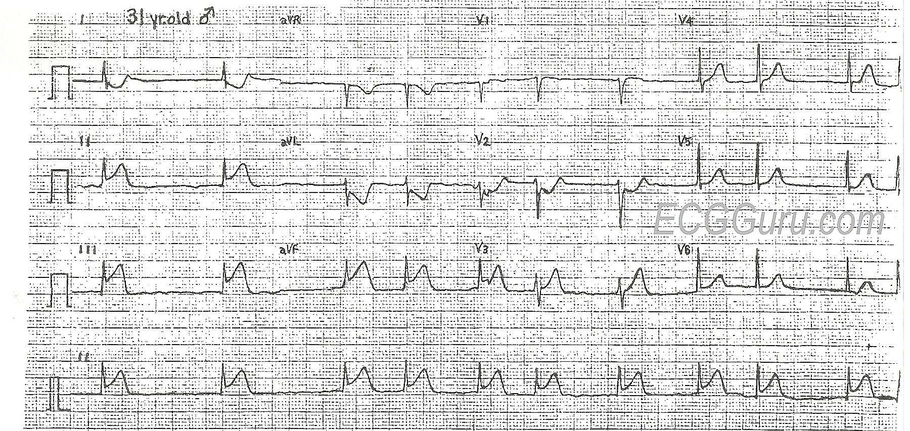
Acute Inferior Wall M.I. With Right Ventricular M.I. and Atrial Fibrillation

This 31-year-old man presented to the Emergency Dept. complaining of chest pain, shortness of breath, and nausea. His heart rate on admission was 120 - 130 bpm and irregular, and the monitor showed atrial fibrillation. His rate slowed with the administration of diltiazem. His 12-lead ECG shows the classic ST elevation of inferior wall M.I. in Leads II, III, and aVF. This patient also had JVD, bibasilar rales, orthopnea, and exertional dyspnea, signs of CHF. He had no history of acute M.I., CHF, or atrial fibrillation. He offered no history of drug use or medications.

This ECG is very useful for the basic student, in that the ST elevations are readily seen, and the atrial fib is definitely irregularly-irregular. For the more advanced student, the ST depression in V2 indicates posterior wall injury, while the flat ST segment in V1 indicates a possible right ventricular M.I.  While the posterior wall is trying to depress the ST segment, the right ventricle is trying to elevate it, resulting in flattening. Also, Lead III has a greater STE than Lead II, which has been shown to be a reliable indicator of RV infarction.  This should be confirmed with a V4 right, or all chest leads done on the right side. Right ventricular injury has been shown to increase mortality, and it also requires different management of hemodynamics.

It is unusual for a 31-year-old to experience acute M.I.  That makes it important to rule out other causes of ST elevation and chest pain.  Benign early repolarization and pericarditis should be considered.  Some of the ECG signs that FAVOR the diagnosis of STEMI are:  1) ST segments are straight, rather than curved downward like a smile.  2)  ST elevations are seen in related leads - leads oriented over the inferior wall and right ventricle (II, III, aVF, V1).  3) Reciprocal ST depressions are seen in leads known to be reciprocal to the inferior leads (I, aVL) and leads reciprocal to the "upper" inferior wall, or posterior wall.  4) There is an acute dysrhythmia (atrial fib).  Atrial fibrillation is a fairly common complication of acute M.I., and also leads to increased mortality, especially when associated with CHF.


All our content is FREE & COPYRIGHT FREE for non-commercial use

Please be courteous and leave any watermark or author attribution on content you reproduce.