The Patient:A 75-yr-old man called Emergency Services because of chest pain and shortness of breath. He had just returned from a 15-mile bicycle ride, during which he had to stop several times to catch his breath, not normal for him.He was so diaphoretic, they were not able to get a good 12-lead ECG. While the paramedics were assessing him and preparing for transport, he went into ventricular fibrillation. He was defibrillated at 360 joules within seconds of onset, and converted to sinus rhythm with pulses.
The ECGs:
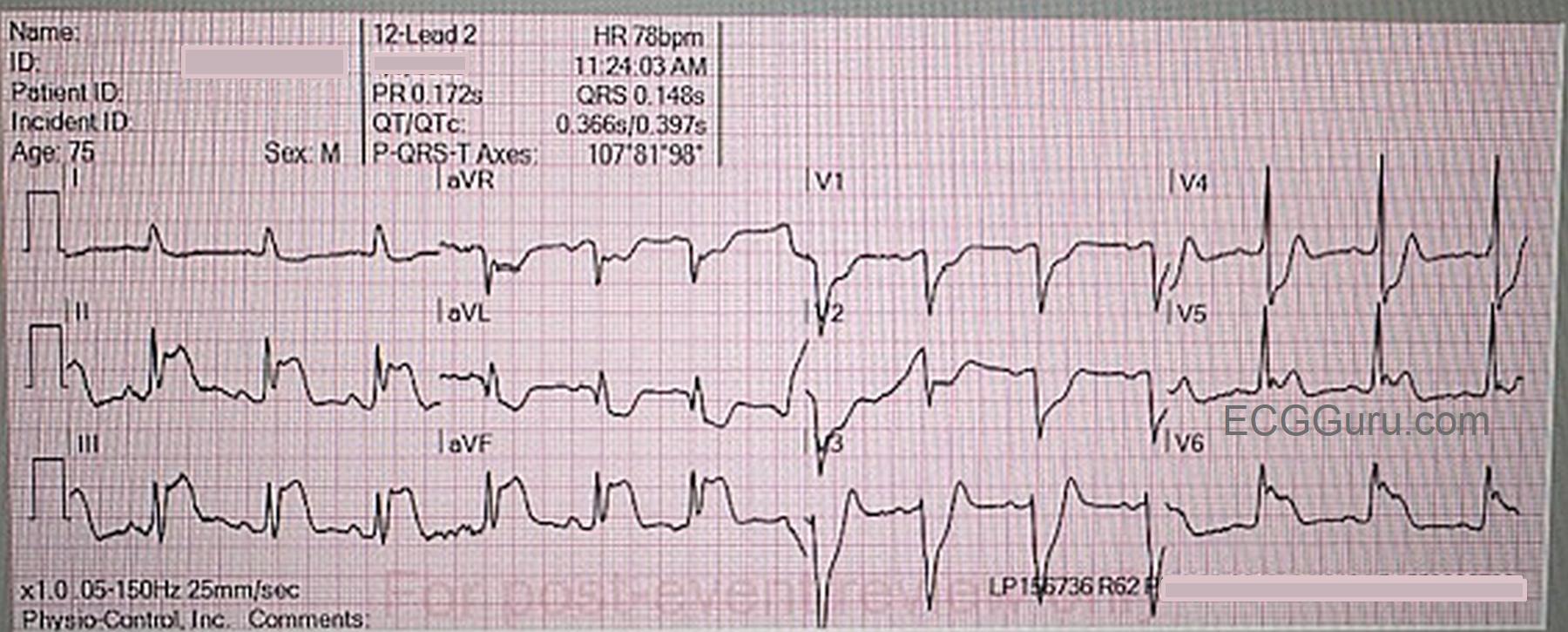
11:24 am: This ECG is obtained after defibrillation, and there is return of spontaneous circulation (ROSC). The rhythm is sinus at 78 bpm. The QRS is slightly wider than normal at .12 seconds. (It is my opinion that the ECG machine read the QRS wider because of the J point and ST changes.) Other intervals and frontal plane axis are within normal limits. R wave progression is normal.
There is marked ST elevation in Leads II, III, & aVF, with reciprocal ST depression in Leads I, aVR, & aVL. (inferior wall transmural injury). There is ST elevation in V5 and V6 (low lateral transmural injury), and ST depression in V1 through V4 (posterior-lateral transmural injury with reciprocal changes in these leads.)