Displaying 1 - 2 of 2
Dawn's picture

Severe Triple Vessel Disease

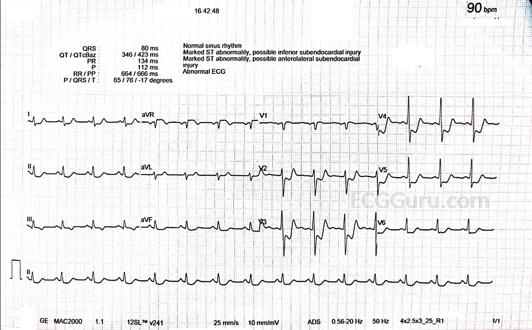
The Patient:   This ECG is from a 63-year-old man who complained of epigastric pain for three hours. The pain was sudden in onset, burning in nature, and accompanied by nausea and palpitations.  The patient is a heavy smoker, diabetic and hypertensive with a long history of non-compliance to his medications. 

He was given crushed aspirin, loaded with clopidogrel and heparin, given high-intensity statins, and rushed to the cath lab. 

The ECG:  The rhythm is normal sinus, a bit fast at 90 bpm.  The intervals, frontal plane axis, and R wave progression are normal.  This ECG shows a very dreaded pattern:  ST segment elevation in aVR and V1 with widespread ST depression, seen here in all other leads.  This is an ECG sign of GLOBAL ISCHEMIA.  There are several possible causes, all bad.  The most common causes of this pattern are:

·        Severe triple vessel disease, with significantly decreased flow in the left anterior descending, right, and circumflex arteries.


Dawn's picture

ST Elevation in aVR and Proximal Arterial Occlusion

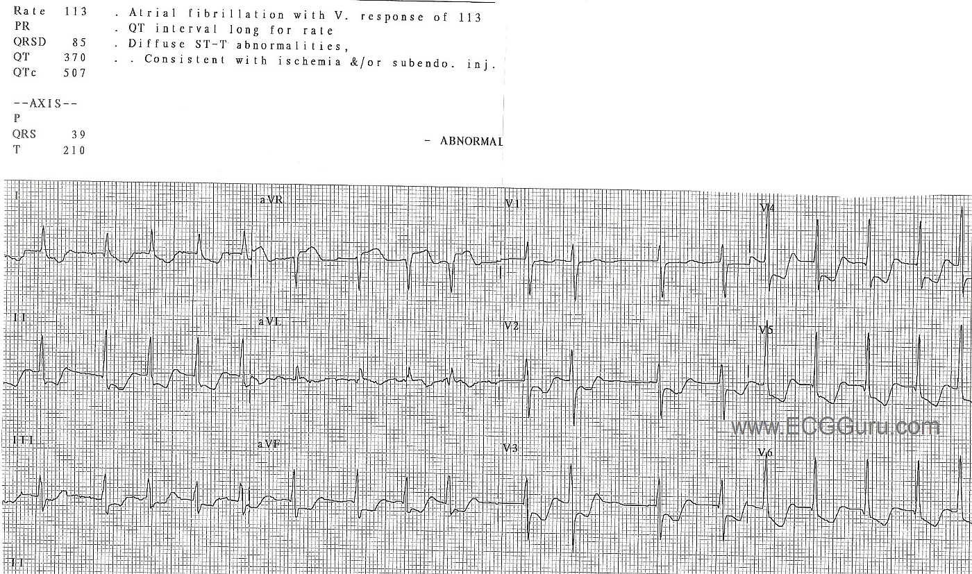
This ECG is from a patient with chest pain.  We have no other information about the patient.  The rythm is atrial fibrillation with a ventricular response of about 113/min.  There is  ST elevation in aVR, with ST depression in all other leads, except V1 and aVL.  V1 is probably caught between showing us ST elevation from the base of the heart (like aVR) and ST depression from the anterior wall (like V2).  Therefore, the ST is flat.  aVL is often elevated in proximal LCA lesions, as well.

This pattern in a person with chest pain is associated with severe acute coronary syndrome (ACS), often with conditions that cause cardiogenic shock.  Proximal left coronary artery obstruction or severe triple vessle disease are likely.

All our content is FREE & COPYRIGHT FREE for non-commercial use

Please be courteous and leave any watermark or author attribution on content you reproduce.