Displaying 1 - 10 of 12
Dr A Röschl's picture

Atrial Fibrillation With Rate-related Left Bundle Branch Block

For a better overview, the leads aVL and V2-V4 are not shown in this ECG. The basic rhythm is atrial fibrillation (no P waves or flutter waves visible, but fibrillation waves). When the conduction rate drops, the QRS complexes are narrow. Faster conduction results in wide QRS complexes with LBBB morphology. This is an example of phase 3 (acceleration dependant) LBBB.

Dawn's picture

Atrial fibrillation and Type 2 M.I.

The Patient:     This excellent teaching case was donated to the ECG Guru by our friend, Sebastian Garay (who is an ECG Guru himself).  It was taken from a 33-year-old man who was complaining of chest pain and palpitations. He reported a similar episode about six months prior, but failed to follow up with cardiology. Was told by his medical care provider that he had atrial fib.

The ECG:      We are able in this case to provide a 12-lead ECG with each lead recorded for the entire width of the paper. This has the advantage of producing twelve ten-second rhythm strips.  Page one contains the limb leads, and page two shows us the precordial leads.

The rhythm is atrial fibrillation, with a heart rate of 133 bpm and an irregularly irregular rhythm. The QRS axis is extreme left at about -75 degrees.  This has caused Leads II, III, and aVF to be negatively deflected, and aVR and aVL to be positive. Lead I is biphasic, low voltage, and mostly positive, indicating that the axis travels almost perpendicular to Lead I, but slightly toward it.

The machine mistakenly gives us a reading for PR interval and P wave axis, even though there are no P waves.  The QRS is on the wide side without being abnormal at .10 seconds (100 ms). The QTc is within normal limits, although it might be considered “borderline”, with 431-450 usually considered borderline.

Dawn's picture

Inferior Wall M.I. With Atrial Fibrillation

The Patient   This ECG was obtained from a 74-year-old man who had a history of COPD. He was complaining of severe chest pain at the time of the ECG.

The ECG     The rhythm is atrial fib or flutter (the R to R intervals are irregular, but seem to repeat about 4 interals).  Flutter waves are seen during some of the longer intervals. The rate is approximately 90 beats per minute.  The ST segments are very noticeably elevated in Leads II, III, and aVF.  There is reciprocal ST depression in Leads I and aVL, and also in all the precordial leads. 

Dawn's picture

ECG Basics: Onset of Atrial Fibrillation

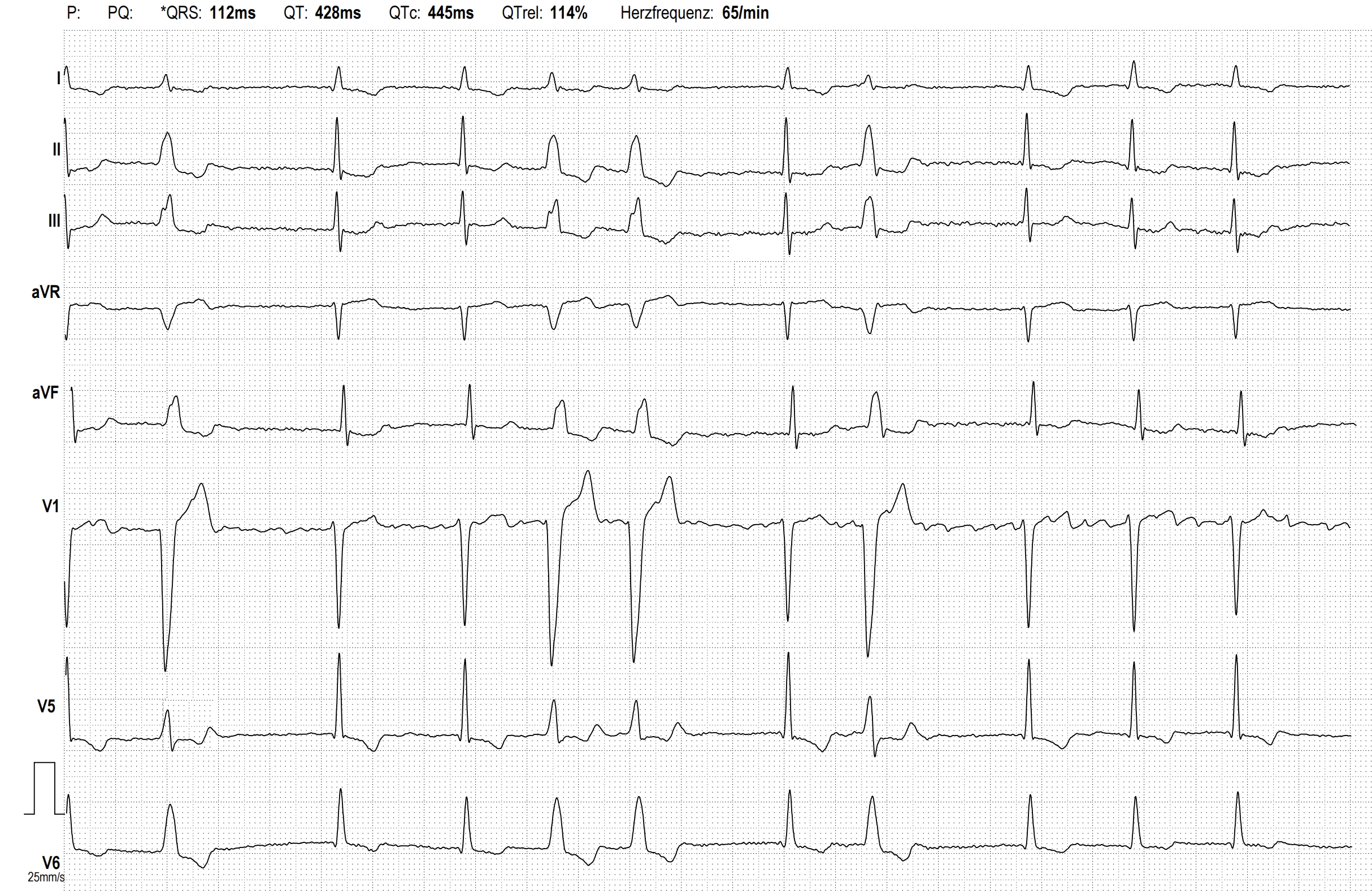
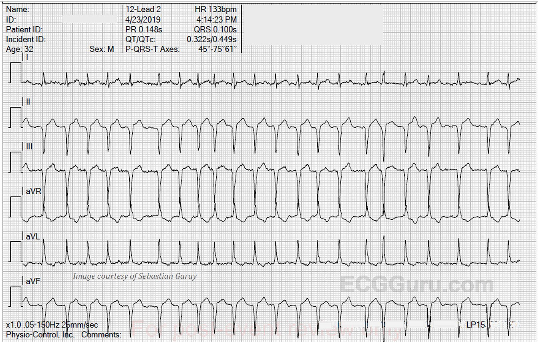
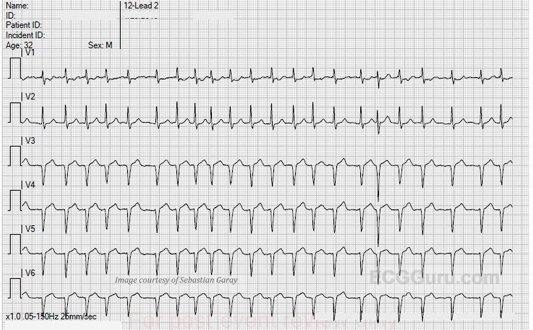
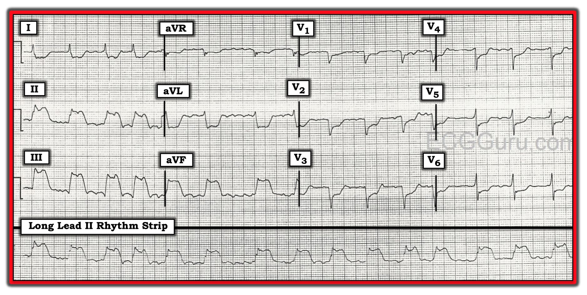
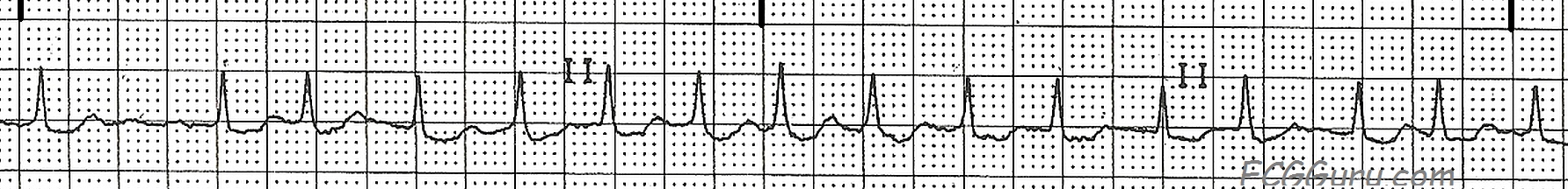
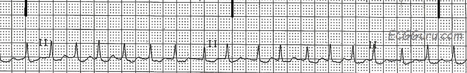
This strip shows the onset of atrial fibrillation.  A fib can be "paroxysmal," meaning that it has a sudden onset, but then stops spontaneously, usually within 24 hours to a week.  A fib can also be classified as "persistent", meaninging that the a fib lasts more than a week.  It can stop spontaneously, or be halted with medical treatment.  "Permanent" a fib is a fib that is resistent to treatment.  

The first three beats in this strip represent sinus rhythm at 75 beats per minute.  At the onset of atrial fibrillation with beat number four, the rhythm becomes irregularly irregular, and the rate is around 140-150 bpm. We can expect new-onset a fib to have a fast ventricular rate, as the atria are sending hundreds of impulses to the AV node every minute. The AV node will conduct as many of those impulses as it can to the ventricles.  Most AV nodes can easily transmit 130-160 bpm.  In a fib, the atria are quivering, not contracting. Because of this fibrillation of the atrial muscle, a fib has no P waves, and therefore, no "atrial kick".  The contribution of the atria to cardiac output (25-30%) is lost. An extremely fast rate can also lower output and overwork the heart, so one treatment goal for a fib is to lower the rate.  This can be done independently of attempts to convert the rhythm.

During a fib, blood clots can form in parts of the atria, especially the left atrial appendage.  If sinus rhythm is restored after these thrombi form, they can embolize and travel to the brain, causing stroke.  Before electively converting atrial fib to a sinus rhythm, the patient may need to be anticoagulated.


Dawn's picture

ECG Basics: Atrial Fibrillation With A Rapid Ventricular Response

This ECG rhythm strip has all the hallmarks of atrial fibrillation:  the rhythm is irregularly irregular and there are no P waves.  The rate is about 150 beats per minute. There is no P wave because the atria are being irregularly depolarized by many ectopic pacemakers at once, causing the atria to "quiver".  This patient has new-onset atrial fib, and has been medicated with a calcium channel blocker.  The rate shows signs of slowing, but has not reached the target rate for this patient of less than 80 bpm.

At the onset of atrial fib, the rate is usually fast, because the AV node is being bombarded by numerous impulses from the atria.  The impulses arrive irregularly, and with different "strengths".  The AV node conducts as many impulses as it is able to, usually resulting in a rate over 110-120 bpm.  Medications can affect the rate, of course, and we use medications to slow AV conduction and allow a more normal heart rate.  

There are many methods of correcting atrial fib, not always with permanent success. Some patients tolerate this rhythm well as long as the rate is kept in check.  But others suffer a loss of cardiac output due to the loss of "atrial kick", which is the forceful filling of the ventricles by the contracting atria.  This loss of cardiac output can severely impair some people, making it necessary to try to convert the atrial fibrillation.  In addition, people living with atrial fib must be anticoagulated, as the loss of forceful emptying of the atria can cause collections of blood clots which can break free and embolize.

Dawn's picture

ECG Basics: Atrial Fibrillation With a Rapid Ventricular Response

This rhythm strip is recorded in two simultaneous leads, which is always preferable to one single lead.  It is a good example of atrial fibrillation with a rapid ventricular response.  Atrial fib that has not been treated will usually have a rapid ventricular rate.  This reflects the ability of the AV node to conduct a tachycardia, within limits.  The natural slow conduction of the AV node allows it to act as a "filter", preventing the huge numbers of impulses generated by the atrial fibrillation from reaching the ventricles.  In this case, about 140 beats per minute are able to make it through the AV node into the ventricles.   In some patients, preexisting cardiac conditions such as valve insufficiency or CHF may make this rate dangerous for the patient.  The rate may lower cardiac output in some people, and this must be considered in light of the fact that the loss of P waves in atrial fib also lowers cardiac output significantly.

Dawn's picture

ECG Basics: Atrial Fibrillation With Rapid Ventricular Response

This is a good rhythm strip to use to illustrate how atrial fibrillation can almost look regular when the rate is fast.  Students should be taught how to "march out" the rhythm for regularity.  It is always a good idea with atrial fib to take a longer strip, looking for the inevitable "gaps" in the R-to-R intervals.  Also, as your students progress, a 12-lead ECG is invaluable to really search for P waves.  Even two or three simultaneous leads are better than just one.

Dawn's picture

ECG Basics: Atrial Fibrillation With A Controlled Ventricular Response

Atrial fibrillation with a controlled ventricular response is often considered to be atrial fib that has been controlled with medication.  While this is often true, it is possible to see atrial fib with a ventricular rate between 50 and 100 bpm in a patient who has not been treated.  The hallmark signs of atrial fib are:  no P waves and irregularly-irregular rhythm.

Multiple, simultaneous leads are advised to be sure there are no P waves, and a long rhythm strip is often needed to prove the irregularly-irregular rhythm.  Remember, even though this rate is adequate (about 90 bpm), the loss of P waves means no "atrial kick".  The atria are not filling the ventricles with a forceful pumping action. Patients can lose a significant percentage - up to 25% - of their cardiac output because of this.  Coupled with the risk of stroke from the formation and embolization of blood clots from the sluggish circulation in the atria, it is much preferable to have sinus rhythm at the same rate.

Dawn's picture

ECG Challenge From Dr. Ahmed - Patient With Hyperkalemia

This ECG was kindly donated to the ECG Guru website by Dr. Ahmed for open discussion among our members.  The patient was a 70+ year-old man who presented with a complaint of dizziness.  His serum potassium level was found to be 6.5 (normal is 3.5 - 5.0).  Upon correction of his K levels, his rhythm was atrial fibrillation at 130 / min.

WHAT DO YOU THINK ABOUT THIS INITIAL ECG?  What is causing the slow rate? Is there atrial activity?  Do you see QRS morphology changes?  What about the anterior ST and T wave changes? 

We look forward to hearing from our members!  And thanks to Dr. Ahmed for donating this interesting ECG.


Dawn's picture

ECG Basics: Atrial Fibrillation With Rapid Ventricular Response

This is a good basic rhythm strip example of atrial fibrillation with a rapid ventricular response showing the identifying characteristics of atrial fibrillation:  no P waves, an irregularly-irregular rhythm, and a "fibrillatory" baseline.  The wavy baseline will not be seen in all leads in all patients, so it is best to use the first two findings as diagnostic criteria.  Atrial fib often appears initially as a rapid rhythm, as the AV node is being bombarded by many impulses from multiple foci (pacemakers) in the atria.  Depending upon the AV node's ability to transmit these impulses,however, we could see a slow, normal, or rapid ventricular response. 

Atrial fib has very chaotic depolarization of the atrial muscle, resulting in quivering and ineffective pumping of the atria.  This loss of "atrial kick" can severely reduce ventricular filling, and can reduce cardiac output by as much as 25%.  In patients with a very rapid rate, cardiac output can be further reduced, causing CHF.  In addition, the fibrillating atria can form blood clots due to sluggish movement of blood.  These clots can embolize and cause stroke.  For these reasons, patients with atrial fib are anticoagulated and sometimes the atrial fib is stopped by medical, surgical, or electrical therapy.  Recurrence of atrial fib is common after treatment, and for some patients, control of the ventricular rate and anticoagulation become the preferred treatment.

Pages

All our content is FREE & COPYRIGHT FREE for non-commercial use

Please be courteous and leave any watermark or author attribution on content you reproduce.