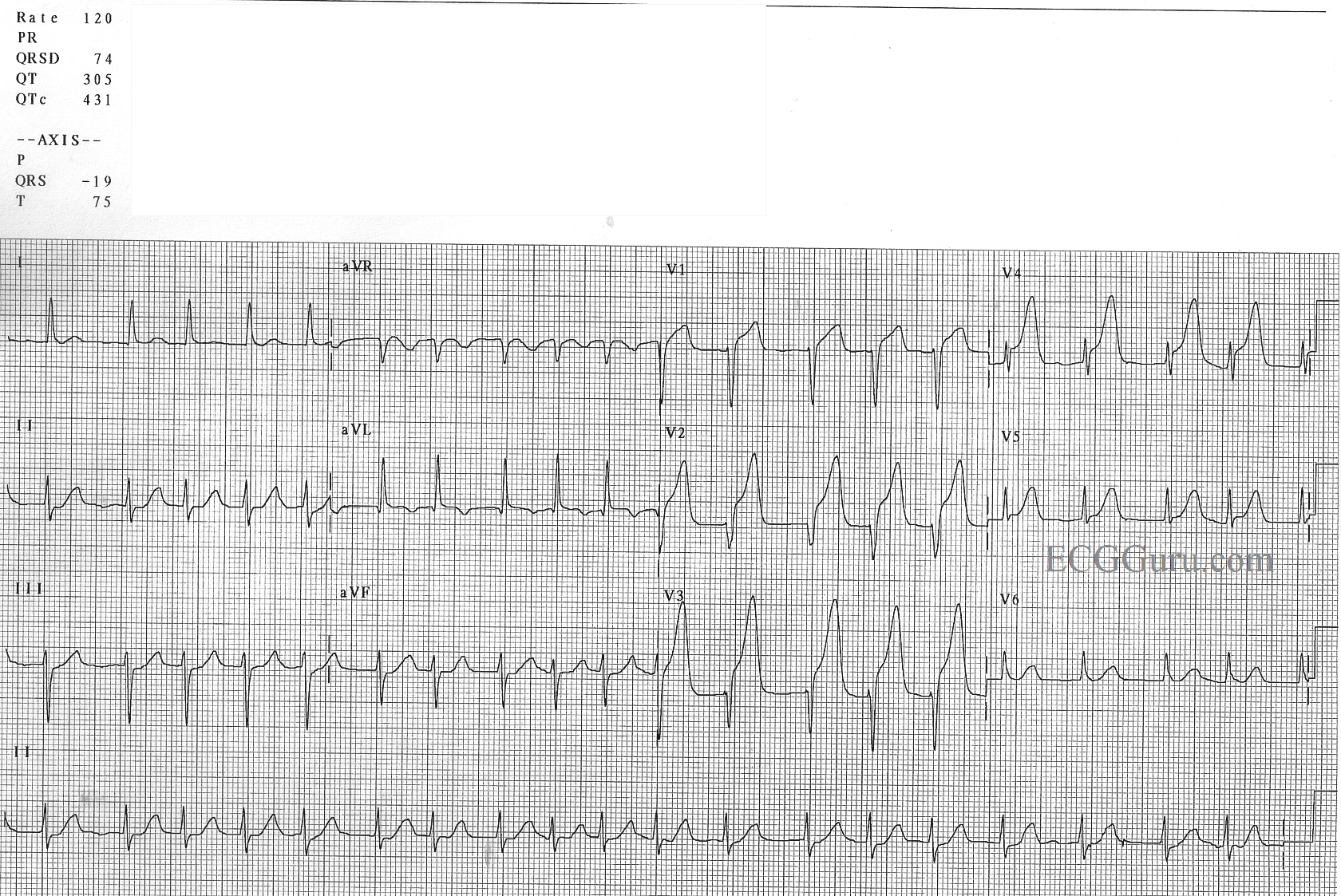
A good teaching ECG, showing clearly elevated ST segments in V1 through V4. This patient had an LAD occlusion. In addition, he has new-onset atrial fib at a rate of about 120/min. Atrial fib has decreased cardiac output because of the loss of P waves prior to the QRS complexes, and a resultant decreased ventricular filling pressure. In addition, this fast rate contributes to increased myocardial oxygen demand and damage. An early priority, along with getting this patient to PCI, is slowing the rate.
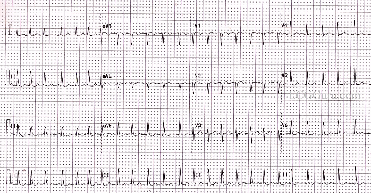
This is a good example of atrial fibrillation with a rapid ventricular response. The rate is around 150 per minute, and the rhythm is almost regular. Show your students how to "march out" the QRS complexes so they can see the irregularity. When the rate is around 150, also check for atrial flutter with 2:1 conduction. In this case, none of the 12 leads shows strong evidence of flutter waves, but it is a possibility when the rhythm is almost regular. Fortunately, the initial treatment for both AFib and AFlutter with rapid response are the same.
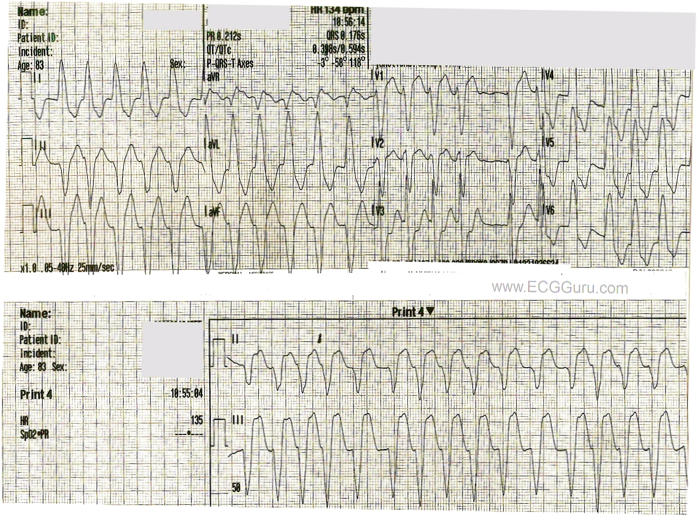
This is a good example of wide complex tachycardia that must be evaluated for V Tach vs supraventricular rhythm with left BBB.
There is an irregular rhythm. When the rate is fast, it is important to look at a longer strip, as sometimes fast rates will cause the rhythm to look regular. We know that monomorphic V Tach is not irregular, so that tells us that we are looking at atrial fibrillation.
The ECG criteria for left bundle branch block is met here: 1) supraventricular rhythm (atrial fib), 2) wide QRS, and 3) negative QRS in V1 and positive QRS in Leads V6 and I.
With wide complex tachycardia, there is always a chance of ventricular tachycardia, and the patient should be treated as V tach until proven differently. For more on determining whether a WCT is V tach or SVT with ventricular conduction delay, go to this LINK.