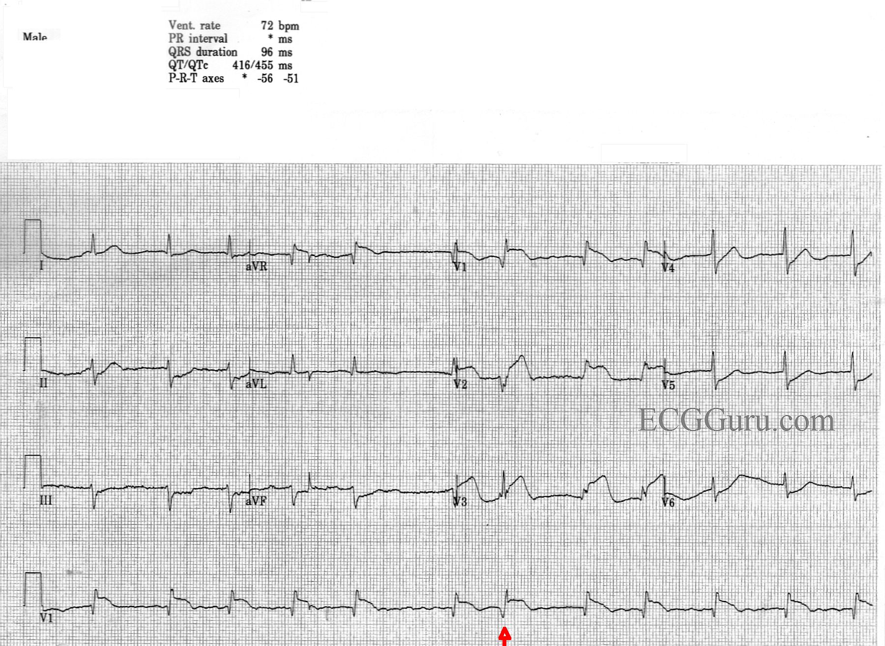
This is an interesting teaching ECG on many levels. It is obtained from a man with chest pain. No other history or follow up is available.
Acute M.I. Most striking is probably the clearly-seen anterior-septal wall M.I.There is ST segment elevation in Leads V1, V2, and V3, with ST depression in the low-lateral leads, V5 and V6.There is also ST depression in the inferior Leads II, III, and aVF.The ST elevations have a coved-upward (frown) shape in V1 and a straight shape in V2 and V3.Both of these ST shapes are abnormal and reflect injury.The depressions are presumed to be due to reciprocal changes, since there is no other ST-depression producing condition apparent.There are abnormal Q waves in V1, which could herald the onset of pathological Q waves, a sign of necrosis, in the anterior-septal wall.