Displaying 1 - 4 of 4
Dawn's picture

ECG Basics: Second-degree AV Block, Type II

This rhythm strip was obtained from a man who was suffering an acute inferior wall M.I.  There are ST elevation and hyperacute T waves.  The rhythm is SINUS ARRHYTHMIA WITH SECOND-DEGREE AV BLOCK, TYPE II.    There is also first-degree AV block.

There are more P waves than QRS complexes, with a 3:2 ratio.  The atrial rate varies between 55 -68 beats per minute.  The sinus rate speeds slightly after the dropped QRS in each group. The ventricular rate is about 40 bpm, with grouped beating. (Regularly irregular.)

The PR intervals are steady at 226 ms (slightly prolonged).

Dawn's picture

ECG Challenge: Guillain-Barre' Syndrome Patient

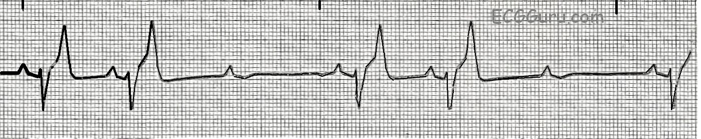
This ECG is probably not for the basic ECG interpretation class.  But, it presents a challenge for the experienced ECG Gurus and instructors out there.  We will leave it here for one week, to allow for comments.  On June 22, we will publish Dr. Jerry Jones’s comments. 

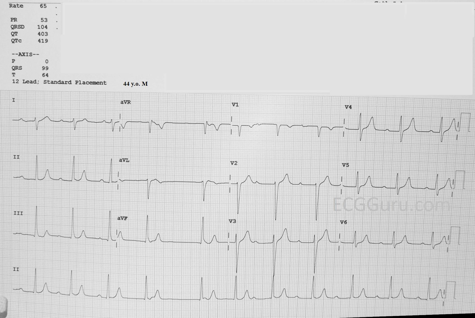
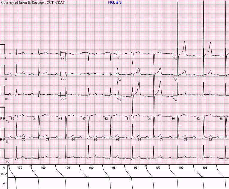
The Patient      This ECG is from a 44-year-old man. He was stricken with Guillain-Barre’ Syndrome when he was 32.  He doesn’t know what his ECGs showed when he was hospitalized with GBS. He knows of no abnormal lab results except for a high CK of 414, attributed to the muscle wasting with GBS.

When he was 43, he started having occasional light-headedness, and was found to have bradycardia around 50 bpm that did not increase with exercise.  A loop recording showed occasional bradycardia over the next several years.  This ECG is now five years old, and the patient says he no longer suffers from bradycardia or lightheadedness, only occasional palpitations and a sensation of “skipped beats”. He lives an active life, albeit with some residual lower extremity weakness from the GBS.

In order to comment on this ECG, it is necessary to “sign in” with an email address.  This is so we can attempt to keep Spammers off the site.  We do not use the email addresses or share them, and we will not contact you. We are looking forward to reading your comments.

Dawn's picture

Irregular Rhythm in a Young Person

The Patient     This ECG is taken from a teenage boy whose mother was concerned that he has an “irregular pulse”.  He is otherwise very healthy, and has no complaints.

The ECG     The rhythm is irregular.  With such a short strip (12-Lead ECGs are often about ten seconds long), it is difficult to determine the exact reason for the irregularity.  One possibility is a sinus rhythm with PACs, which would not be unexpected in a young person.  The more likely interpretation is sinus arrhythmia, also very common in young people.  In a longer strip, we might be able to associate the rhythmic slowing and speeding of the rate with the patient’s respirations.  The P waves all look alike in each of the twelve leads. Different P wave morphology in the early beats would indicate PACs, but the similar P waves do not exclude PACs.

The PR interval is normal, the QRS duration normal, the QTc is normal.  The rate (average 68 bpm) is normal.  The frontal plane QRS axis is normal, with Lead II having the tallest QRS in the frontal plane (limb leads), and aVL small and biphasic.  R wave progression in the precordial leads is good, with the transition from negative to positive in V3. 

All our content is FREE & COPYRIGHT FREE for non-commercial use

Please be courteous and leave any watermark or author attribution on content you reproduce.