Displaying 1 - 10 of 16
Dr A Röschl's picture

Impending Trifascicular AV Block

Here we see the EKG of a 63-year-old man with CAD without relevant coronary stenosis. He complains of slightly reduced performance, but no other symptoms. The ECG shows the following changes:

Dawn's picture

Ask The Expert

Today's Expert is Dr. Jerry Jones, MD, FACEP, FAAEM                                                                                                                                                             

 Jerry W. Jones, MD FACEP FAAEM is a diplomate of the American Board of Emergency Medicine who has practiced internal medicine and emergency medicine for 35 years.    

Dr. Jones has been on the teaching faculties of the University of Oklahoma and The University of Texas Medical Branch in Galveston. He is a published author who has also been featured in the New York Times and the Annals of Emergency Medicine for his work in the developing field of telemedicine. He is also a Fellow of the American College of Emergency Physicians and a Fellow of the American Academy of Emergency Medicine and, in addition, a member of the European Society of Emergency Medicine. 

 Dr. Jones is the CEO of Medicus of Houston and the principal instructor for the Advanced ECG Interpretation Boot Camp and the Advanced Dysrhythmia Boot Camp.                                                                                                                                                                                                                                                                                                                    


Question:  I teach beginner students. How can I explain the complex subject of “AV Blocks”?  I don’t want to teach incorrect information while trying to simplify the subject.


 Answer:  AV Blocks Article By Dr. Jerry Jones  (click link)


Dawn's picture

ECG Basics: Second-degree AV Block, Type II

This rhythm strip was obtained from a man who was suffering an acute inferior wall M.I.  There are ST elevation and hyperacute T waves.  The rhythm is SINUS ARRHYTHMIA WITH SECOND-DEGREE AV BLOCK, TYPE II.    There is also first-degree AV block.

There are more P waves than QRS complexes, with a 3:2 ratio.  The atrial rate varies between 55 -68 beats per minute.  The sinus rate speeds slightly after the dropped QRS in each group. The ventricular rate is about 40 bpm, with grouped beating. (Regularly irregular.)

The PR intervals are steady at 226 ms (slightly prolonged).

Dawn's picture

High-grade AV Block With Profound Bradycardia

If you are an ECG instructor, you probably carefully choose ECGs to illustrate the topic you are teaching. One of the reasons for the existence of the ECG Guru website is our desire to provide lots of such illustrations for you to choose from.

Sometimes, though, an ECG does not clearly illustrate one specific dysrhythmia well, because the interpretation of the ECG depends on so many other factors.  In order to get it “right”, we would need to know information about the patient’s history, presentation, lab results, or previous ECGs. We might need to see the ECG done immediately before or after the one we are looking at.  Some ECG findings must ultimately be confirmed by an electrophysiology study before we can know for sure what is going on.

For those of us who are “ECG nerds”, it can be fun to debate our opinions and even more fun to hear from wiser, more advanced practitioners about their interpretations.

My belief, as a clinical instructor, is that we must teach strategies for treating the patient who has a “controversial” ECG that take into account the level of the practitioner, the care setting, and the patient’s hemodynamic status.  In some settings, it might be absolutely forbidden for a first-responder to cardiovert atrial fibrillation, for example.  But atrial fib is routinely cardioverted under controlled conditions in hospitals.  The general rule followed by emergency providers that “all wide-complex tachycardias are v tach until proven otherwise” has no doubt prevented deaths in situations where care providers did not agree on the origin of the tachycardia.

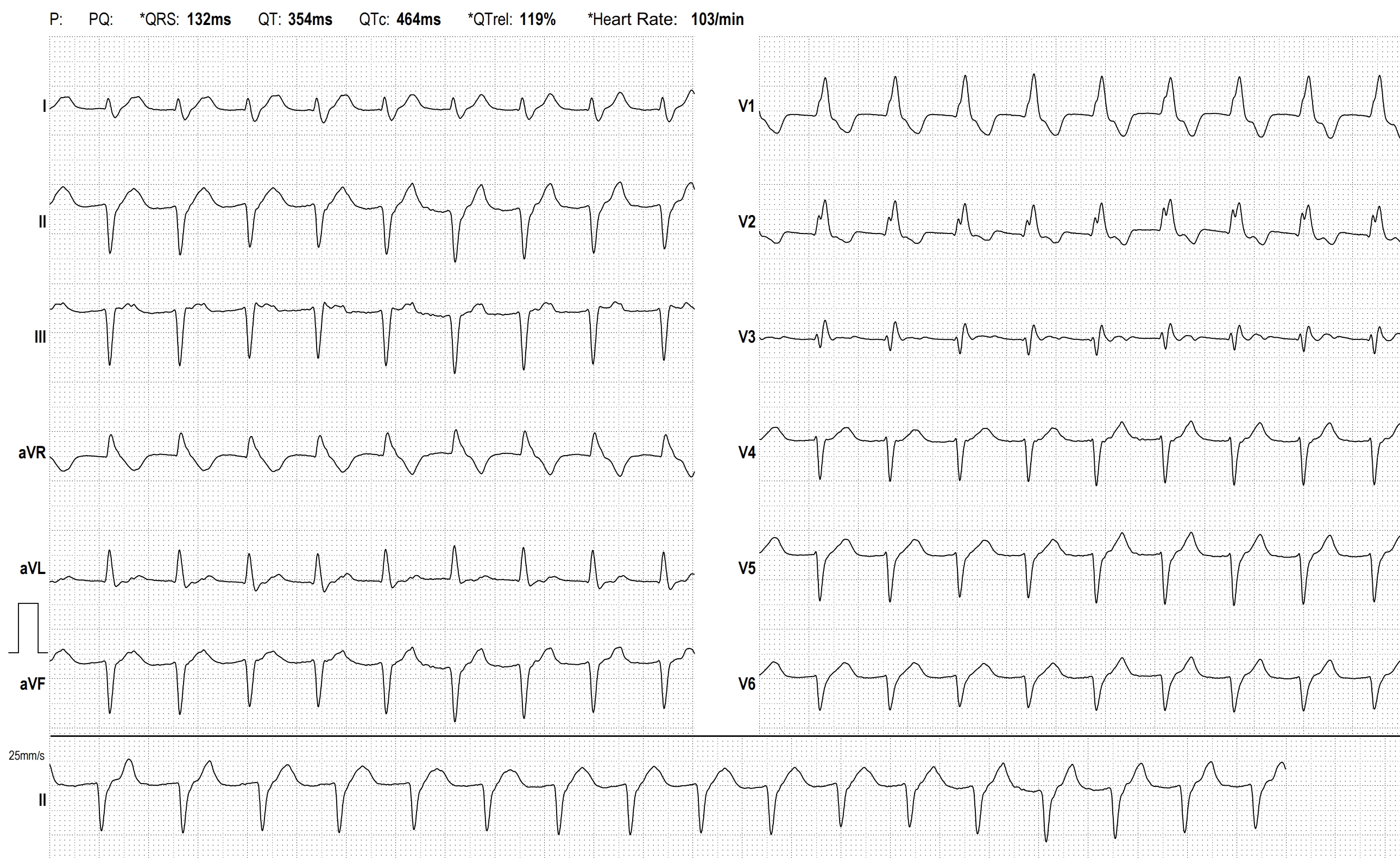
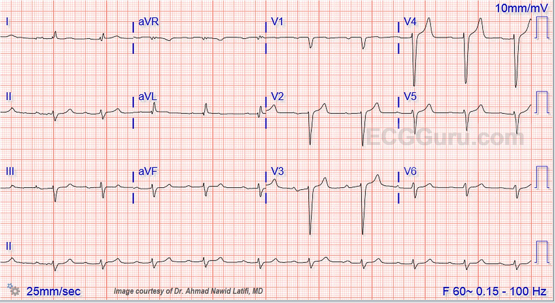
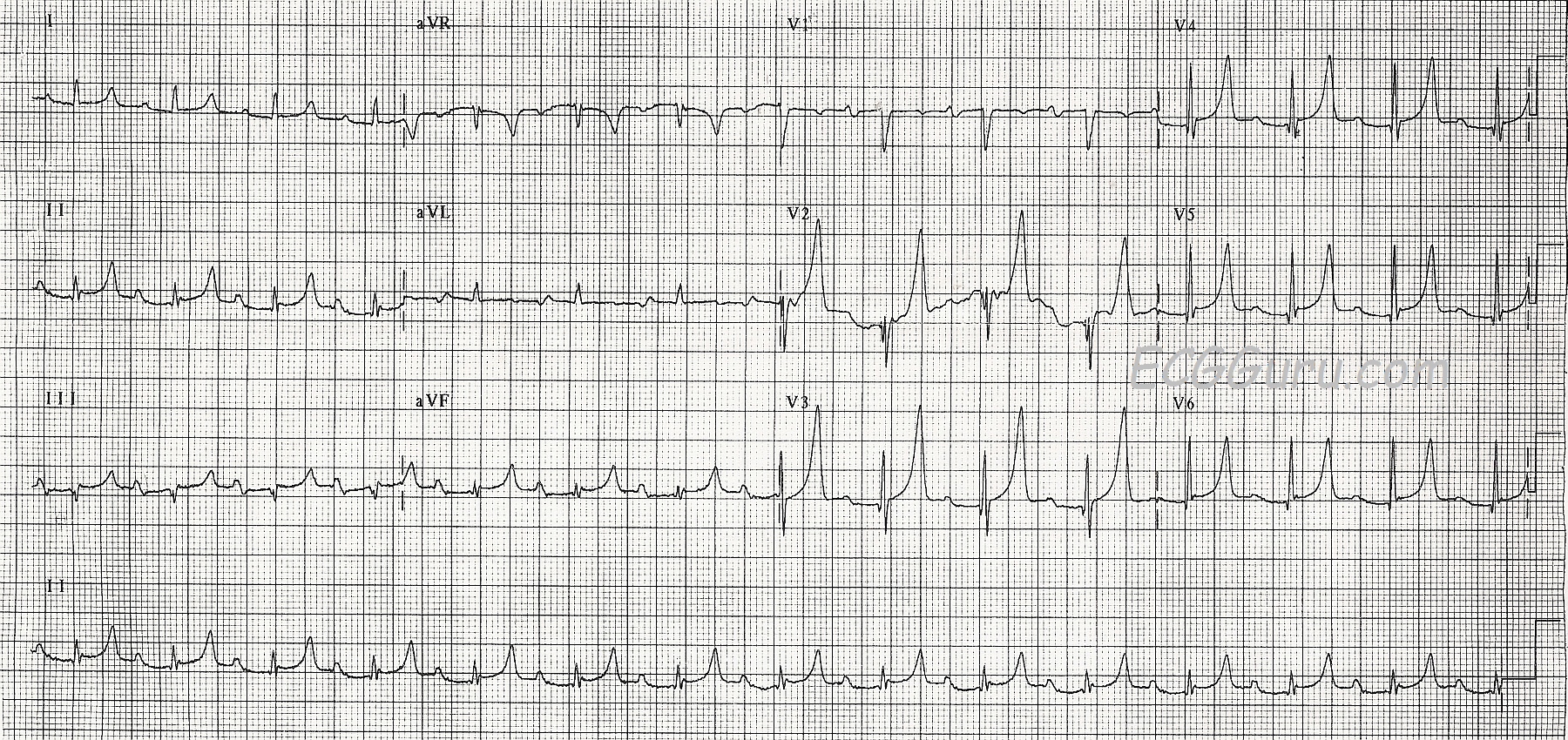
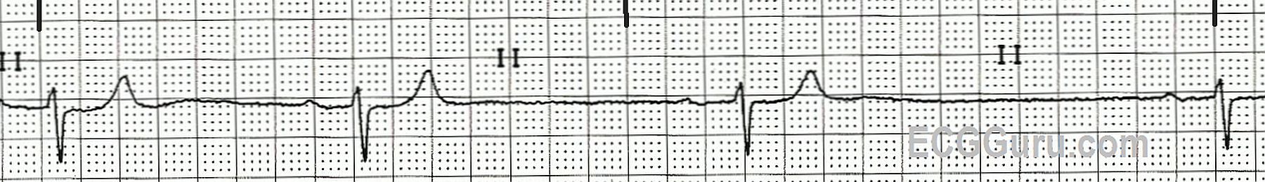
The ECG:    We do not have much patient information to go with this ECG, just that it is from a 71-year-old woman who developed severe hypotension and lost consciousness, but was revived with transcutaneous pacing.   Here is what we do know about this ECG:

·        There are regular P waves, at a rate of about 39 bpm (sinus bradycardia).

Dawn's picture

Previous Anterior Wall M.I.


If you are an instructor, or a fairly new student, you don’t always need to see “challenging” ECGs. But, you may not want to see “standard” ECGs from an arrhythmia generator, either.  Every ECG contains subtle and not, so subtle characteristics of the person it belongs to.  Take a minute to look at this ECG before reading the discussion, and ask yourself what you might surmise about the patient.

The Patient: We don’t know much about the actual patient this ECG came from.  What we do know is that he is an elderly man with a history of heart disease who was hospitalized sometime in the past with an acute M.I.  He is now on beta blocker medication and is on a diet, as he is approaching the “morbidly obese” classification.  He is now in the ER with shortness of breath and mild chest pain.  What does his ECG tell us?

Dawn's picture

Wide QRS Complex With First-degree AV Block

The Patient:  This ECG was taken from a 73-year-old man with a history of heart failure with preserved ejection fraction, severe left ventricular hypertrophy, Type II diabetes, and stage 4 chronic kidney disease.  He also suffered deep vein thrombosis and is on anticoagulation.  He has a recent diagnosis of IgA myeloma.  He presented with a complaint of nausea and vomiting and was found to have a worsening of acute kidney infection.  There was suspicion of renal and cardiac amyloidosis, but the patient refused biopsy to confirm this.  He was started on chemotherapy for multiple myeloma and will be followed as an outpatient.

The ECG:  The rhythm is sinus at around 60 bpm, although the rate varies a little at the beginning of the strip.  The QRS complex is wide at .12 seconds, or 120 ms., representing interventricular conduction delay (IVCD).  The PR interval is .32 seconds, or 320 ms. This constitutes first-degree AV block.  There is left axis deviation in the frontal plane and poor R wave progression in the horizontal plane.

Dawn's picture

Right Bundle Branch Block With Probable Previous M.I.

This ECG was obtained from an 87-year-old man with chest discomfort.  We have no other clinical information.

ECG Interpretation   The rhythm is regular and fast, with P waves, at 95 beats per minute. So, it is normal sinus rhythm, but the rate is probably not “normal” for this patient.  The P waves are small, and difficult to see.  We suggest Lead I to best view the P waves in this example. This is a good opportunity to teach the value of evaluating rhythm strips in more than one simultaneous lead, as subtle features may not show up well in all leads.  There is a first-degree AV block, with a PR interval of 232 ms.

We see the right bundle branch block (RBBB) pattern: rSR’ in the right precordial leads (with a tiny q wave in V1, which is not typical of  RBBB).  The QRS is wide at 148 ms (.148 seconds).  The R prime (R’) represents the right ventricle depolarizing slightly after the left ventricle.  This terminal delay widens the QRS without affecting the depolarization or contraction of the left ventricle.  This delay can be seen in every lead, but is especially easy to see in Leads I and V6, where there is a wide little s wave.  It is normal for the T waves to be in a direction opposite that of the terminal wave (inverted in Leads V1 and III, for example.)

There is left axis deviation.  The causes of LAD are many.  It is not unusual for people with RBBB to also have a left anterior hemiblock (LAH), also called left anterior fascicular block.  The left anterior fascicle has the same blood supply as the right bundle branch.   LAH causes a frontal plane axis shift – instead of Lead II having the tallest QRS of the limb leads, Leads I and aVL will be the tallest upright QRS complexes of the six limb leads.  Lead II will be very small, or flat, or negative. However, the probability of pathological Q waves in the inferior leads offers a more likely explanation for the leftward axis shift.  The M.I. that would have caused these Q waves is old, as there are no acute ST changes.  It would, of course, help to know this patient’s history.

Right bundle branch block can make evaluating for ST segment elevation a bit tricky.  Occasionally, the terminal delay – especially in Leads III and aVF – can be mistaken for ST elevation.  The J points in this ECG all appear to be at the baseline, with no overt STEMI.


Dawn's picture

Non-Sustained Ventricular Tachycardia

This ECG was obtained from a 45-year-old man who was experiencing palpitations and lightheadedness, which he originally attributed to anxiety. There are short, but frequent periods of ventricular tachycardia, which are self-limiting.  This is called "NON-SUSTAINED VENTRICULAR TACHYCARDIA".  The underlying rhythm is sinus, with a remarkably long PR interval, and at least one episode of failure of the P wave to conduct, making "second-degree AV block, Type II" a possibility. It is difficult to thoroughly evaluate the underlying rhythm because it is not seen very often in this ECG. The rate of the underlying P waves is about 67 bpm.  The PR interval is .40 seconds (400 ms).  The "normal" QRS complexes are slightly widened, at about .10 sec (100 ms), which is typical of Type II AVB.  The ventricular QRS complexes are wide at .16 sec. (160 ms)

To assist you in using this tracing for teaching, we have also supplied a "marked up" version.  The P waves, both visible and hidden, are marked with red lines. The PR intervals are shown in the Lead V1 rhythm strip in green.  And the QRS complexes are numbered.  QRS complexes numbered 2, 3, 6, and 12 are sinus. The P wave AFTER QRS #5 is non-conducted.

To review the differentiation of ventricular tachycardia from supraventricular tachycardia with aberrant conduction, go HERE.

Dawn's picture

Hyperkalemia

This ECG was obtained from a patient who had a serum potassium level of 7.4 mEq/L.  It shows some of the earliest ECG signs of hyperkalemia.  There are tall, sharply-peaked T waves in many leads.  The P waves have not yet widened and lost amplitude, but they will soon flatten out and disappear.  At this level of hyperkalemia, we can expect to see conduction disturbances (first-degree AV block in this case) and bradycardia (not yet). It is a bit surprising that the QRS complexes have not yet widened at this serum K level.    Caution:  hyperkalemia can progress and become life-threatening very quickly.

Potassium is primarily an intracellular electrolyte.  It is necessary for proper electrical functioning of the heart.  Extracellular  serum potassium can rise due to renal failure, or taking potassium supplements, potassium-sparing diuretics, or ACE inhibitors.  Occasionally, serum K levels may be artificially elevated by drawing the blood with too much syringe pressure, or using too small a needle, as the red blood cells can be damaged and release intracellular K into the serum.

ECG signs may vary among people with hyperkalemia, but in general:

Serum K levels of 5.5 mEq/L or greater can cause repolarization abnormalities like tall, peaked T waves.

Dawn's picture

ECG Basics: Sinus Bradycardia With First-degree AV Block

This is a nice teaching strip of a slowing sinus bradycardia that began around 40 bpm, and is slowing.  It is a good example of how the sinus node slows down - there is no abrupt change of rates, rather a change with each R-to-R interval.  There is also a first-degree AV block, reflecting slowing of conduction in the AV node.  The PR interval is slightly variable at .28 sec. to about .32 sec.  This is a good strip to begin talking about treatment of bradycardias with beginner students, as there is no second- or third-degee AVB, but the patient is very likely to be symptomatic now, or very soon.  Atropine would probably improve this rate in a symptomatic patient, but if there is time, a 12-Lead would be a good idea to rule out acute M.I.  Inadvertently raising the rate too much in the injured heart can lead to pump failure, while leaving the patient poorly-perfused in a bradycardia will starve the heart.  A transthoracic or temporary IV pacemaker might be a better choice for some patients because of our ability to choose the rate.

Pages

All our content is FREE & COPYRIGHT FREE for non-commercial use

Please be courteous and leave any watermark or author attribution on content you reproduce.