
Submitted by Dr A Röschl on Mon, 08/28/2023 - 06:24
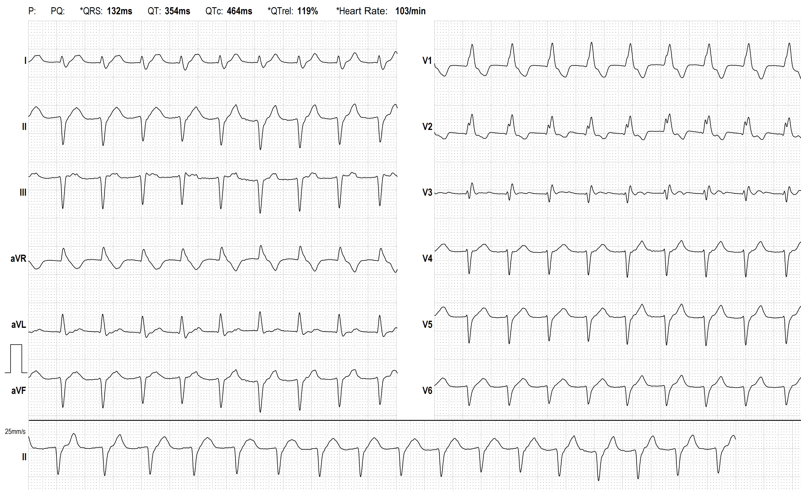
Here we see the EKG of a 63-year-old man with CAD without relevant coronary stenosis. He complains of slightly reduced performance, but no other symptoms. The ECG shows the following changes:
The rhythm is a sinus rhythm with positive P waves in the inferior leads and with a wide distance to the subsequent QRS complex, indicating a long PR interval (approximately 400 ms). The heart rate is 103 beats per minute, thus indicating sinus tachycardia. The P waves are difficult to recognize as they are only seen as small deflections on top of the T waves. In lead V1/V2 the typical pattern of a right bundle branch block is visible (rR' in V1 and V2). An over-rotated left axis type with continuous S waves up to V6 suggests a left anterior fascicular block. Therefore, a bifascicular block pattern is present. Additionally, there is this long AV block I. One can speak of an impending trifascicular block. A pacemaker indication does not arise solely from this EKG finding unless corresponding symptoms are present. Clinical follow-up examinations are of course necessary.
Rate this content:
-

- Dr A Röschl's blog
- Log in or register to post comments
All our content is FREE & COPYRIGHT FREE for non-commercial use
Please be courteous and leave any watermark or author attribution on content you reproduce.