Displaying 1 - 5 of 5
Dr A Röschl's picture

Sinus Bradycardia and More

Let's analyze the ECG. It comes from a pacemaker patient whose pacemaker was briefly switched to VVI at 30 bpm due to a stimulation threshold test. The first 3 beats show a sinus rhythm with a frequency of approx. 40 bpm. This is followed by a premature ventricular contraction (PVC). The P wave of the next sinus node beat lands exactly on the T of the PVC. This cannot be conducted to the ventricles, either because the ventricular myocardium is still unexcitable or the PVC has conducted retrogradely into the AV node and this is therefore still refractory.

Dawn's picture

Anterior Wall M.I. With Bifascicular Block

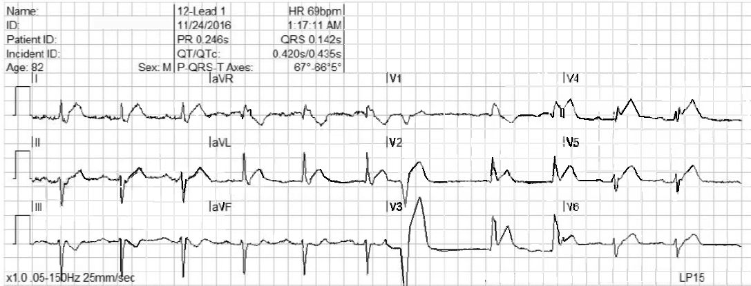
This ECG is taken from an 82-year-old man who called 911 because of chest pain.  He has an unspecified “cardiac” history, but we do not know the specifics. 

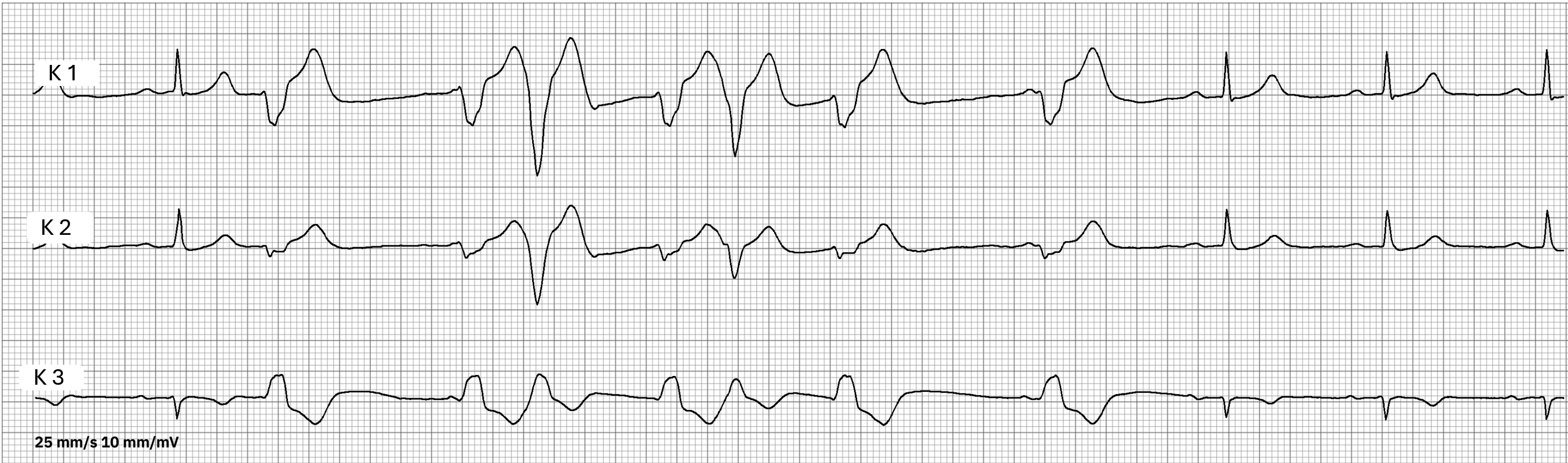
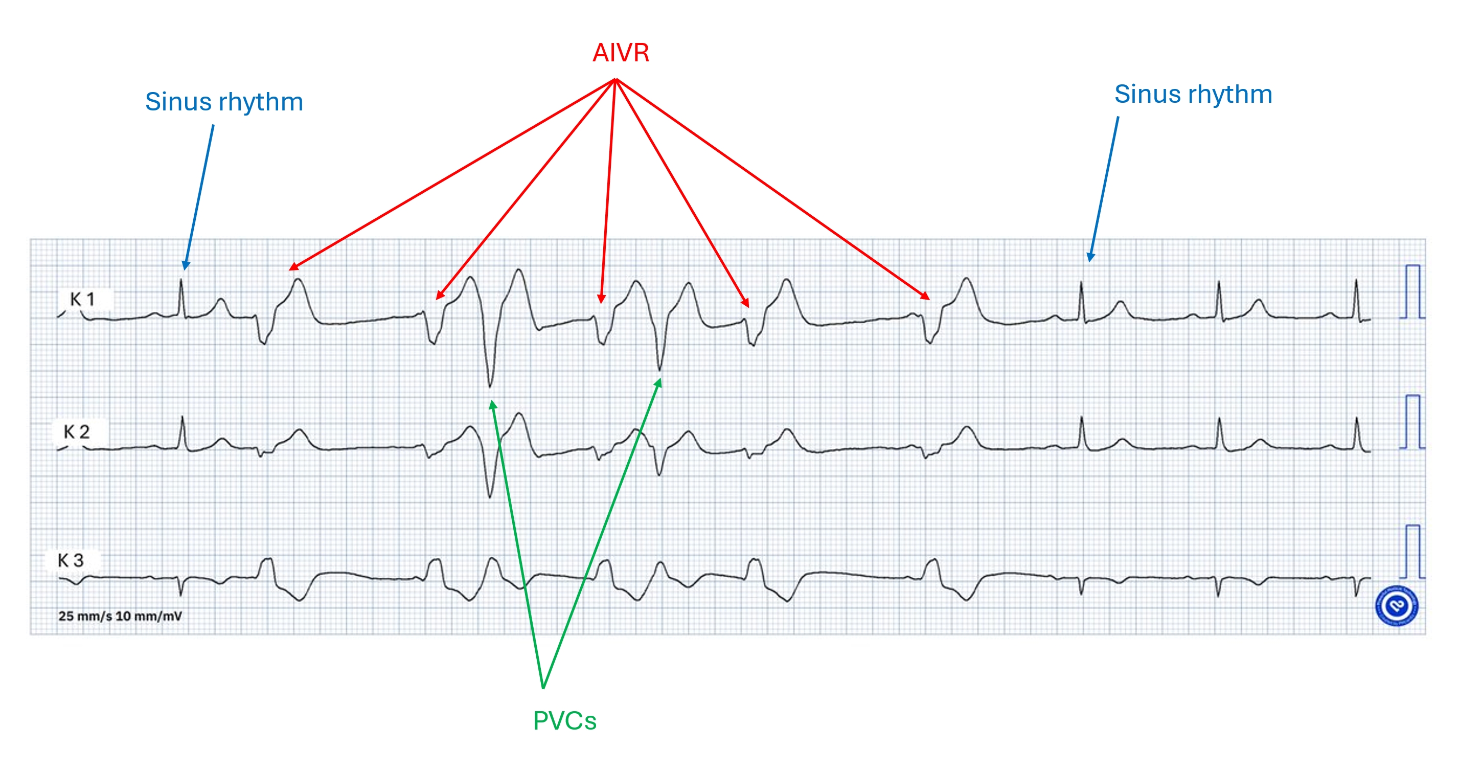
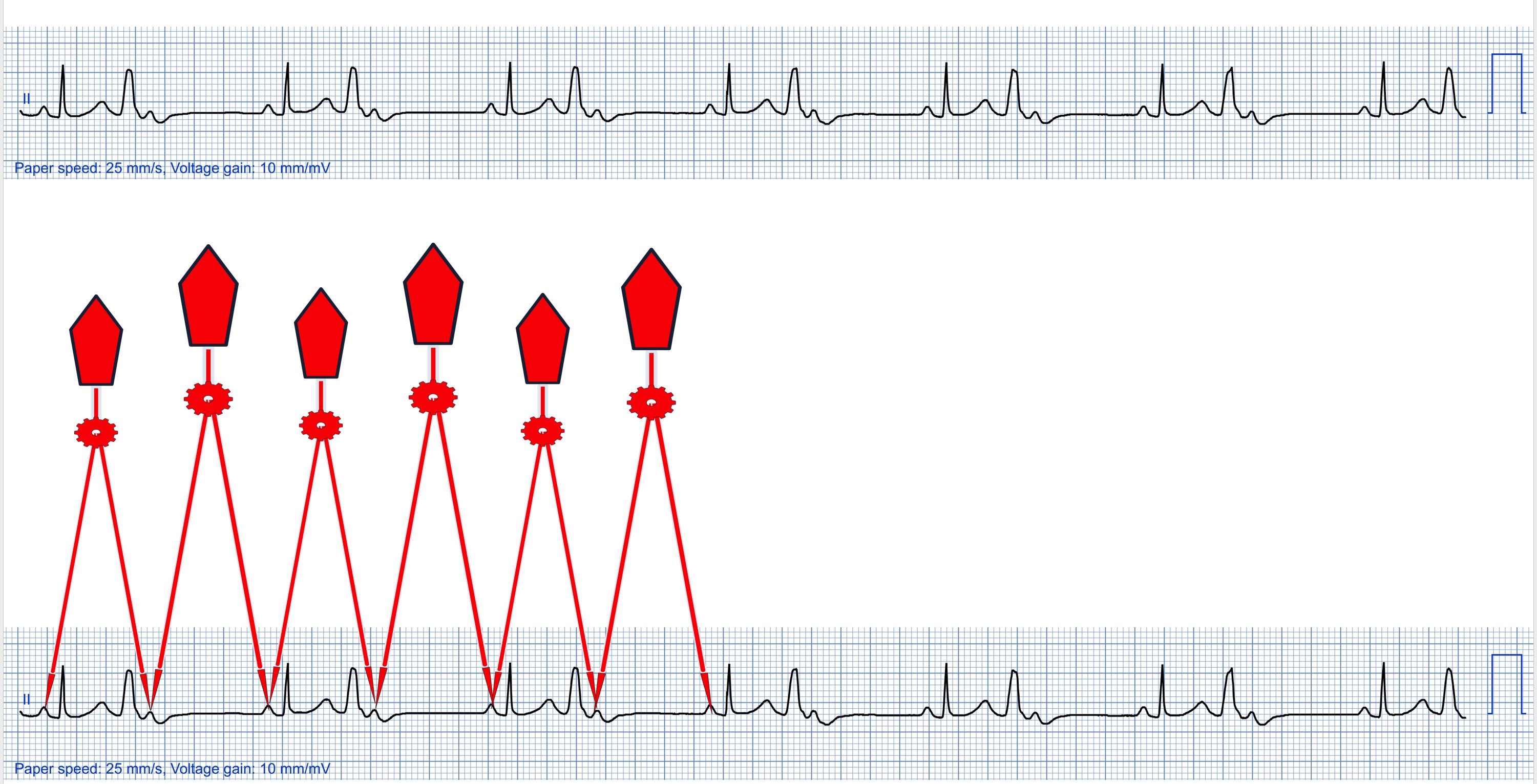
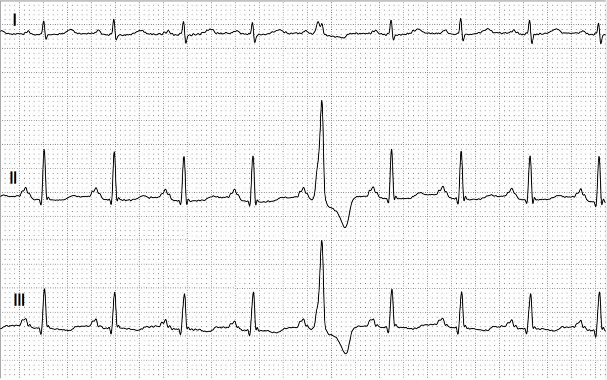
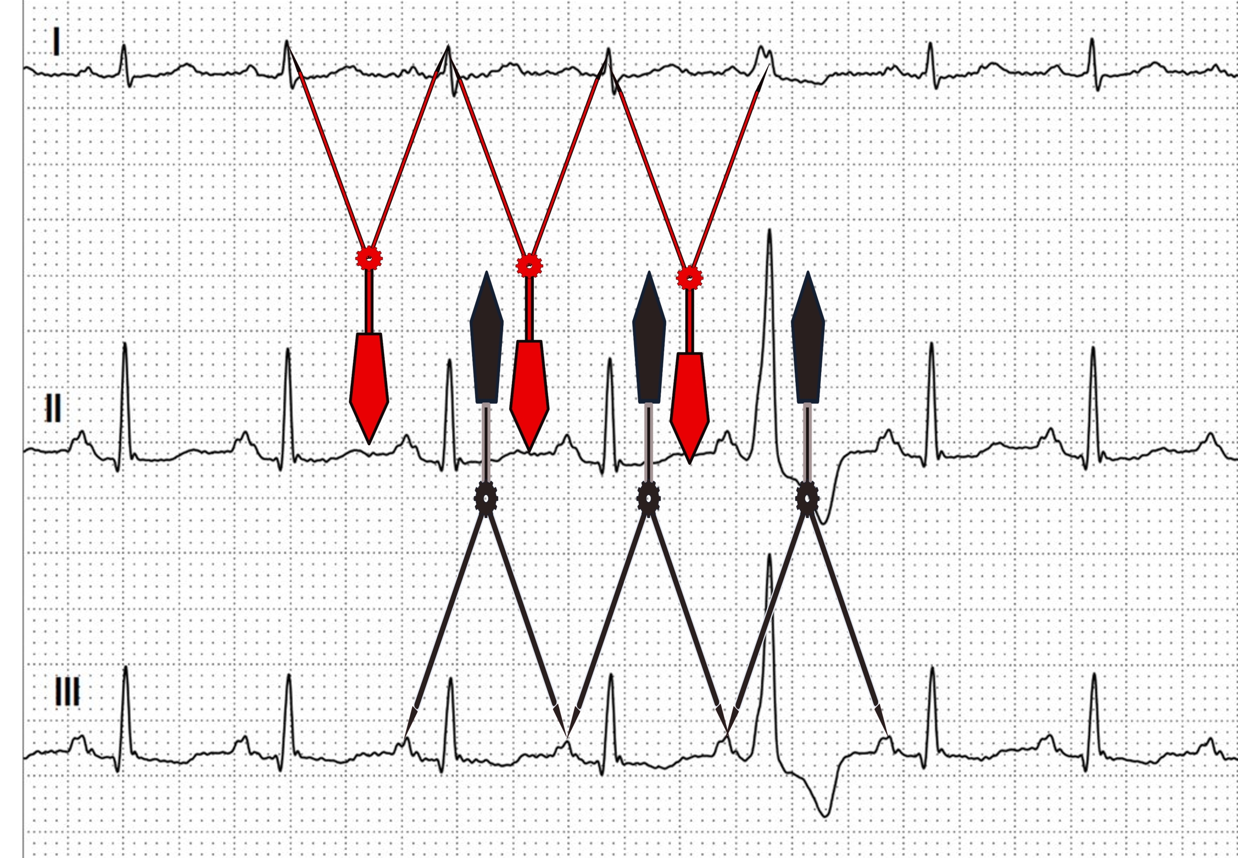
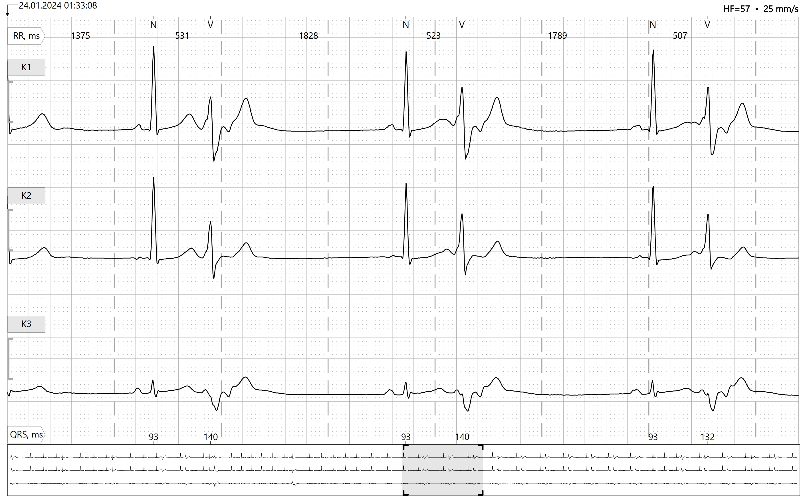
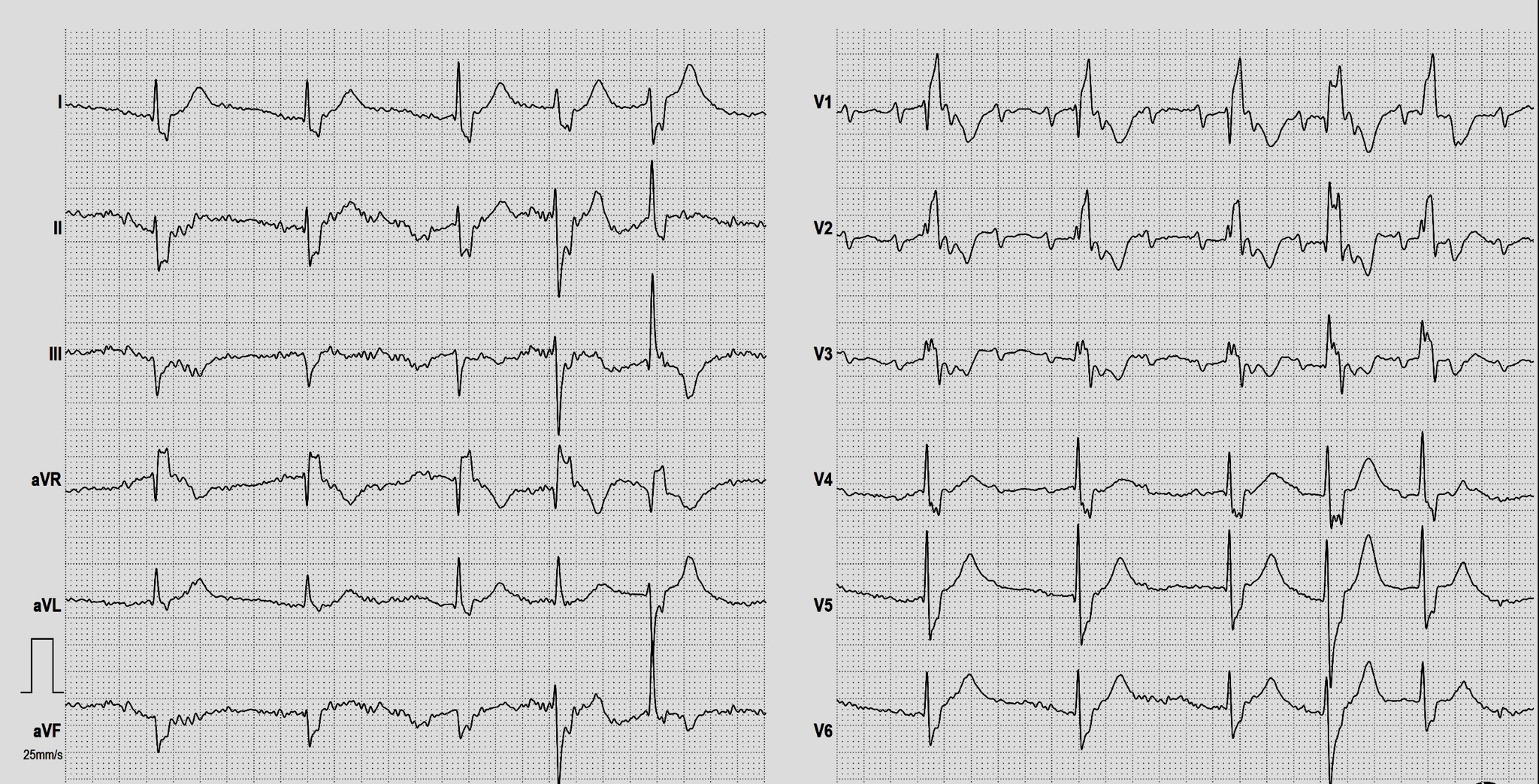
WHAT IS THE RHYTHM?  The heart rate is 69 bpm, and there are P waves before every QRS complex. The underlying rhythm is regular, with one premature beat that is wide without a P wave.  The PR interval is slightly prolonged at .25 seconds.  The rhythm is normal sinus rhythm with first-degree AV block and one PVC. 

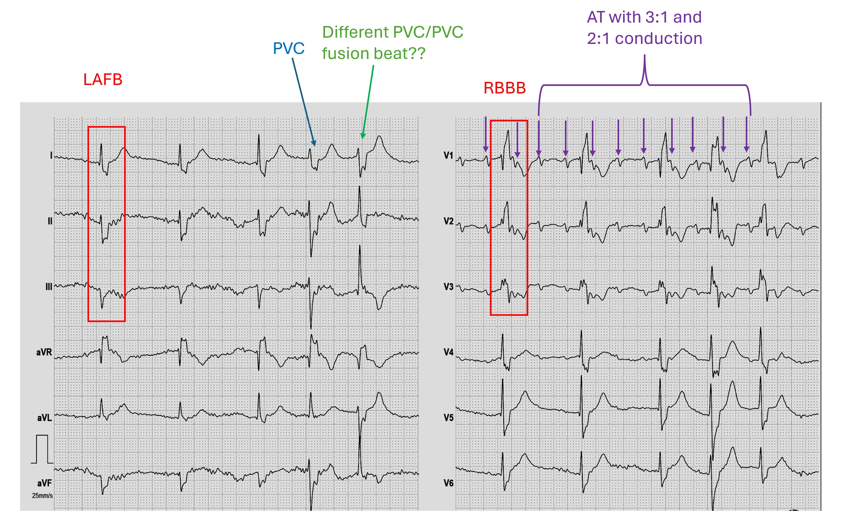
WHY THE WIDE QRS?   The QRS complex is wide at .14 seconds. The QRS in V 1 has a wide R wave after a small Q wave.  This in consistent with right bundle branch block pattern, with loss of the normal initial small r wave (pathological Q waves).  The diagnosis of RBBB is further corroborated by the wide little S waves in Leads I and V6.  The QRS frontal plane axis is -66 degrees per the machine, and clearly “abnormal left” because the QRS in Lead II is negative, while the QRS in Leads I and aVL are positive.  This is left anterior fascicular block, also called left anterior hemiblock.  The combination of RBBB and LAFB is a common one, as the two branches have the same blood supply.  It is also called bi-fascicular block. 

WHAT ABOUT THE ST SEGMENTS?  The ST segments in leads V2 through V6 are elevated, and their shape is very straight, as opposed to the normal shape of coved upward (smile). Even though the amount of ST elevation at the J points appears subtle, the shape of the segments, the fact that they appear in related leads, and the fact that the patient is an elderly male with chest pain all point to the diagnosis of ANTERIOR WALL ST elevation M.I. (STEMI).  Additional ST changes include a straight shape in Leads I and aVL and ST depression in V1 and aVR.  

PATIENT OUTCOME  The patient was transported to a cardiac center, where he received angioplasty in the cath lab.  The left coronary artery was found to be occluded, and was repaired and stented.  He recovered without complications and was sent home in a few days.

Dawn's picture

ECG Basics: Normal Sinus Rhythm With Premature Ventricular Contractions

This ECG shows an underlying rhythm of normal sinus rhythm at a rate of 80 / min.  There are two premature ventricular contractions (PVCs).  The sinus rhythm actually continues uninterrupted, causing a “compensatory pause”.  If you march out the P waves, you may even see hints of the hidden P waves in the ST segments of the PVCs.  The P waves that occur in the ST segments of the PVCs land in the refractory period of the ventricles, and so are unable to continue into the ventricles and cause a QRS. 


It is also permissible to call these beats “ventricular premature beats (VPBs)” or “ventricular premature complexes (VPCs)”.  

Dawn's picture

Sinus Rhythm With Left Bundle Branch Block, PVCs, and Fusion Beats


This is a great ECG for teaching your students about some of the different causes of wide QRS.  This 89 year old man has a sinus rhythm that is around 100 bpm, and his QRS is widened at 148 ms (.148 sec).  Leads I and V6 are positive, and Lead V1 is negative, meeting the criteria for left bundle branch block. There is a left axis deviation, which is common with LBBB, although it is not always this pronounced, indicating that there is possibly another cause for LAD.  In this ECG, there are also PVCs and probable fusion beats.  The 14th beat is a PVC.  Complexes 1, 6, and 9 are possibly fusion beats. Fusion can be described as an almost simultaneous sinus beat and ventricular beat.  The depolarization waves, one coming from the top of the heart and one coming from the bottom, meet and "fuse" on the ECG.  Fusion beats will have some characteristics of the supraventricular beats and some of the ventricular beats.  They are not significant except that fusion can be said to "prove" the existence of a ventricular pacemaker - either a natural pacemaker or an electronic one.

Do you see anything else interesting in this ECG?  How would YOU describe this rhythm?  Please do not hesitate to add your comments, or ask questions of the experts who contribute to this site.  We will respond quickly to all questions.

Dawn's picture

Left Bundle Branch Block In Patient With Severe Aortic Stenosis

This ECG is from a 91-year-old man who was being evaluated for replacement of his aortic valve, which was severely calcified. It shows a classic LBBB pattern: wide QRS, supraventricular rhythm (normal sinus rhythm with first-degree AV block), a negative QRS in V1, and a positive QRS in Leads I and V6.

All our content is FREE & COPYRIGHT FREE for non-commercial use

Please be courteous and leave any watermark or author attribution on content you reproduce.