Dawn's picture

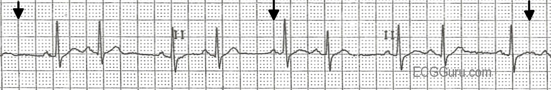
This is a normal sinus rhythm with atrial bigeminy, a term meaning that every other beat is a PAC.  If you look carefully, you can see slight differences in the sinus P waves and the atrial (premature) P waves.  The PACs penetrate and reset the sinus node, causing what looks like a delay after the PAC.  It is often just a return to the normal P to P interval, or nearly so.  If you teach basic students in a clinical setting, they will learn from palpating the peripheral pulse and feeling the pattern of bigeminal beats. Sometimes, the premature beat feels much weaker due to less filling time available to the ventricles.  Atrial bigeminy can have very benign causes, such as increased caffeine intake, or it can have more complex causes such as advanced heart disease or conduction blocks.  In some patients, atrial bigeminy, or any PACs, can be a precursor to more serious atrial dysrhythmias, such as atrial fibrillation.

Rate this content: 
Average: 3.4 (8 votes)

Comments

ekgpress@mac.com's picture

There is "beauty" in the simple lead II rhythm strip presented this week by Dawn. For the beginning interpreter - it is simply sinus rhythm with PACs - here presented in a bigeminal pattern (every-other-beat a PAC).
 
On an intermediate level - We really do NOT know for sure these are "PACs" - since I am not certain P wave morphology is definitely changing ... Although the P wave preceding the 2nd, 6th and 8th beats does appear to be a bit more pointed than P waves preceding sinus beats - the P preceding beat #4 is more rounded and looks virtually identical to other sinus P waves. Same is true for the very last P wave on the tracing (whose QRS is cut off). Given baseline artifact (Note slightly changing morphology for virtually every ST segment) - I do not think one can comfortably say that P wave morphology is in fact changing ... 
 
Additional leads are often needed in order to determine IF P wave morphology is truly changing vs attributing minor differences in P wave morphology to artifact. Imagine a PAC arose in the right atrium from a location very close to the SA node. To lead II - such a PAC would look very much like the P wave of normal sinus beats. It is only with additional leads (on a 12-lead tracing) - that one may be able to appreciate that P wave morphology is in fact truly changing.
 
On an advanced level - the above concept may be more than academic. IF in fact P wave morphology is not changing in this tracing - then the group beating with pause less-than-twice the shortest R-R interval could be consistent with SA block rather than atrial bigeminy.
 
Finally, on a more advanced level - Note QRS morphology of the 4th and especially 6th beats is slightly different than that of the other sinus beats (the QRS looks to be a little shorter). It is possible there may be some element of aberrant conduction with these earlier-than-anticipated beats. That said - given the overal changing P wave and ST-T wave morphology and baseline undulations (see between beats #8-9) - I think it more likely that any changes in QRS morphology seen are artifactual, perhaps from respiratory variation - BUT the advanced interpreter should recognize these slight differences in some of the early beats, and then process whether this effect is likely to be "real" (ie, aberrant conduction) vs artifactual.
  • For those interested - I've abstracted 9 pages from my ACLS-2013-Arrhythmias (Expanded Version) book - that review the above concepts on using additional leads for assessing P wave morphology - as well as laddergram illustration of SA block. CLICK HERE to download this PDF.


Ken Grauer, MD  www.kg-ekgpress.com   [email protected] 

...or is it sinus arrhythmia?

All our content is FREE & COPYRIGHT FREE for non-commercial use

Please be courteous and leave any watermark or author attribution on content you reproduce.