Dawn's picture

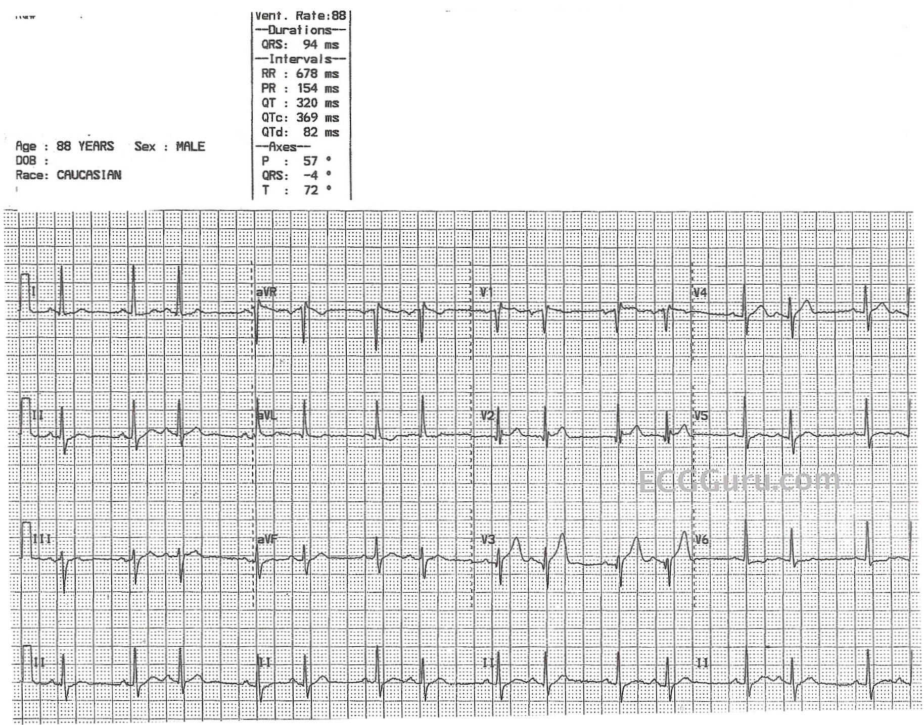
This ECG is from an 88-year-old man with congestive heart failure.  No other clinical information is known.  It shows an underlying sinus rhythm with atrial bigeminy - every other beat is a premature atrial contraction.  There is very little, if any, difference in the morphology of the sinus P waves and the ectopic P waves, indicating that the ectopic focus is in the vicinity of the sinus node.  There is no "compensatory" pause, because PACs penetrate the sinus node, resetting it.  So, the underlying sinus rate here is about 72 beats per minute.  There are several mechanisms for bigeminy to occur, but ectopic bigeminy is the most common.

Rate this content: 
Average: 4.5 (4 votes)

Comments

ekgpress@mac.com's picture

 
     This is an extremely interesting tracing. As per Dawn — there is a bigeminal rhythm. However, I am uncertain as to whether this represents atrial bigeminy, or perhaps SA Block of the Wenckebach type.
 
  • The KEY — and the difficulty — lies with determining IF P wave morphology of sinus beats and of the 2nd beat in each group is the same. If every-other-beat is truly a PAC — then P wave morphology in at least a few of the simultaneously recorded leads on this 12-lead tracing should show differences (even if subtle) in P wave morphology. Instead, I see slight differences in morphology among MANY of the P waves on this tracing, including among the P waves that initiate each of the 7 groups we see in the long lead II rhythm strip at the bottom of the tracing. This baseline “noise” (artifact) prevents me from determining with any certainty if all P waves are coming from the SA node. If they are — then there is Wenckebach periodicity in the form of group beating with duration of the pause being less than twice the shortest R-R interval.
  • PACs are far more common than SA block. That said — SA block does occur, and often goes unnoticed. It should be thought of when there is group beating with sudden drop of a P wave and QRS complex in a setting in which all P waves manifest the same morphology. Unlike sinus pauses which are random and for which the pause is often more than twice the shortest R-R interval — SA Wenckebach typically is repetitive with pauses of similar length that are less than twice the shortest R-R interval.
  • Otherwise — this 88-year old man with heart failure has an abnormal ECG. There is an rSr’ in lead V1 — and Q waves in leads V2,V3. This may represent prior anterior infarction (which may be a substrate for SA block). I suspect there is either an abnormal body habitus and/or precordial lead misplacement given the prominent R wave in V2 that then diminishes unexpectedly in V3 … ST-T wave changes do not appear to be acute.
  • The reason for slight difference in QRS morphology for the 2nd beat in each pair in some leads is aberrant conduction (because of the early occurrence of the 2nd QRS complex in each group).
NOTE: Click here for brief Review of SA Block —


Ken Grauer, MD  www.kg-ekgpress.com   [email protected] 

All our content is FREE & COPYRIGHT FREE for non-commercial use

Please be courteous and leave any watermark or author attribution on content you reproduce.