
Submitted by Dr A Röschl on Fri, 06/02/2023 - 05:47
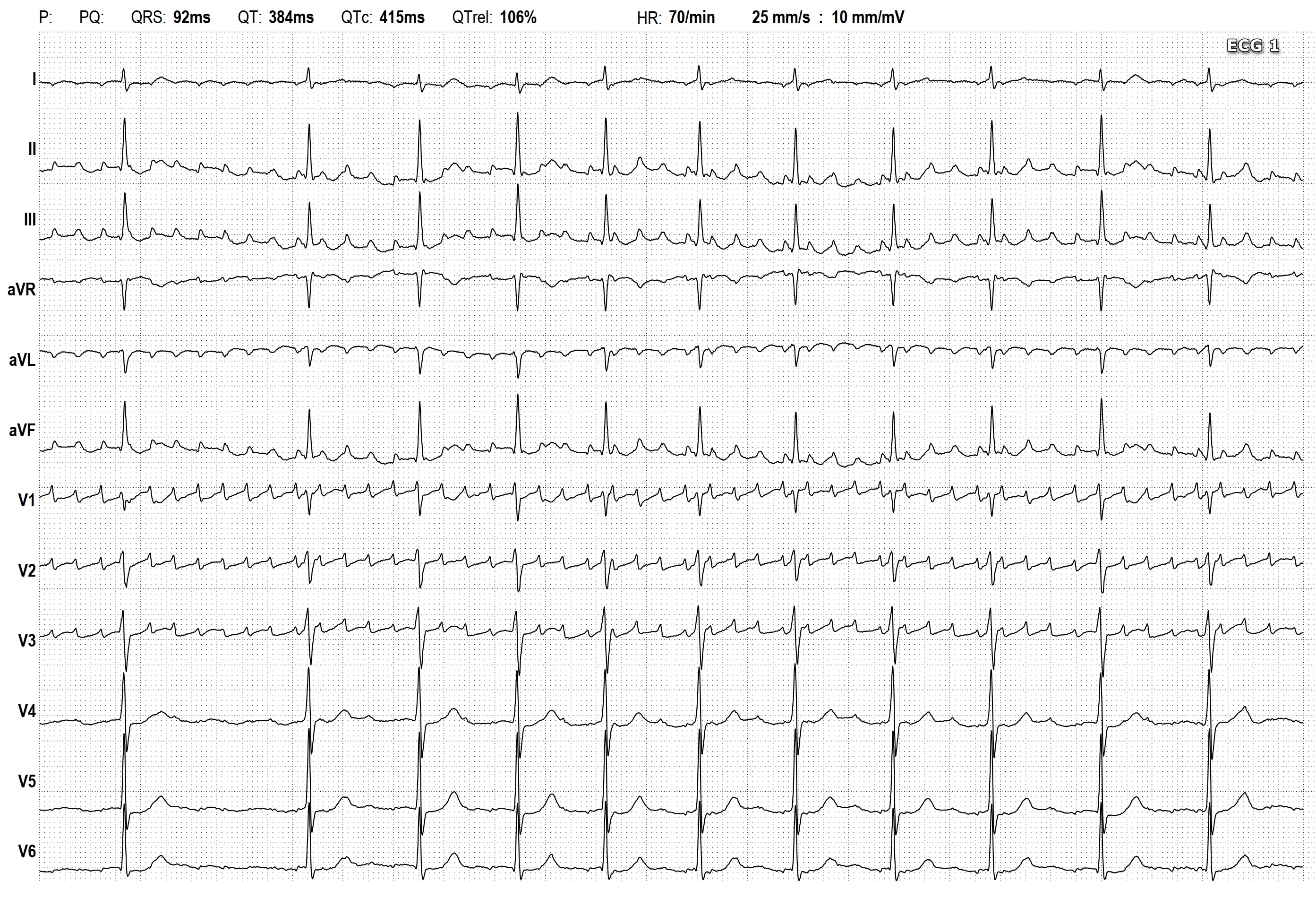
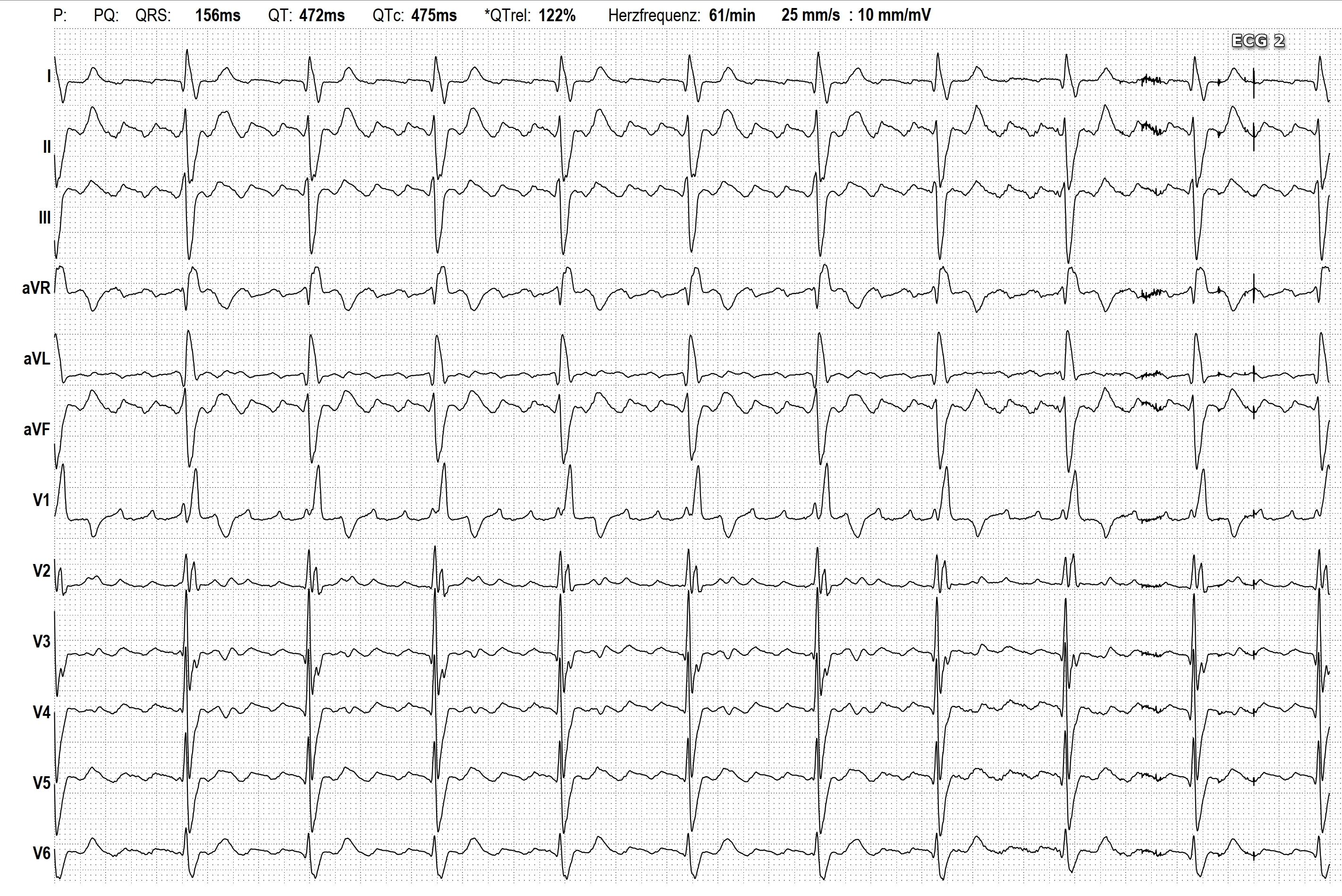
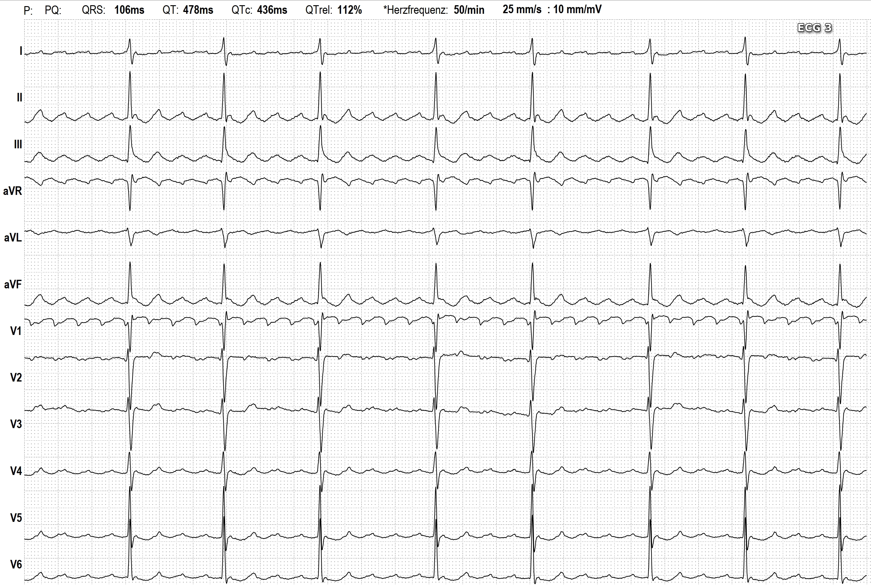
Why is this left atrial atypical atrial flutter (ECG 1)? Atrial fibrillation can be excluded because nice flutter waves (all look the same) can be clearly identified. With typical right atrial flutter, the reentry circle runs counterclockwise and we see typical saw tooth patterns in the inferior leads (negative flutter waves). The flutter waves are positive in V1 (ECG 2). With typical right atrial flutter with a clockwise reentry circle, the flutter waves in the inferior leads are positive. In V1 they are variable and show typically a w-shaped pattern (ECG 3). There is no zeroline between the flutter waves in both forms in the inferior leads (reentry circle!)
In this ECG, showing left atrial atypical atrial flutter, the flutter waves in lead I are negative. Lead I goes from the right to the left arm, with the positive pole on the left arm. So the vector of the lead is opposite to the direction of spread of the flutter waves. So the flutter waves travel from left to right, away from the positive electrode. A zeroline can be seen between the flutter waves in nearly all leads (focal origin!) It is not always possible to discern the different forms of atrial flutter in a 12 lead ECG.
Rate this content:
-

- Dr A Röschl's blog
- Log in or register to post comments
All our content is FREE & COPYRIGHT FREE for non-commercial use
Please be courteous and leave any watermark or author attribution on content you reproduce.