
Submitted by jer5150 on Sun, 10/21/2012 - 14:34
No clinical patient data available for this 12-lead ECG.
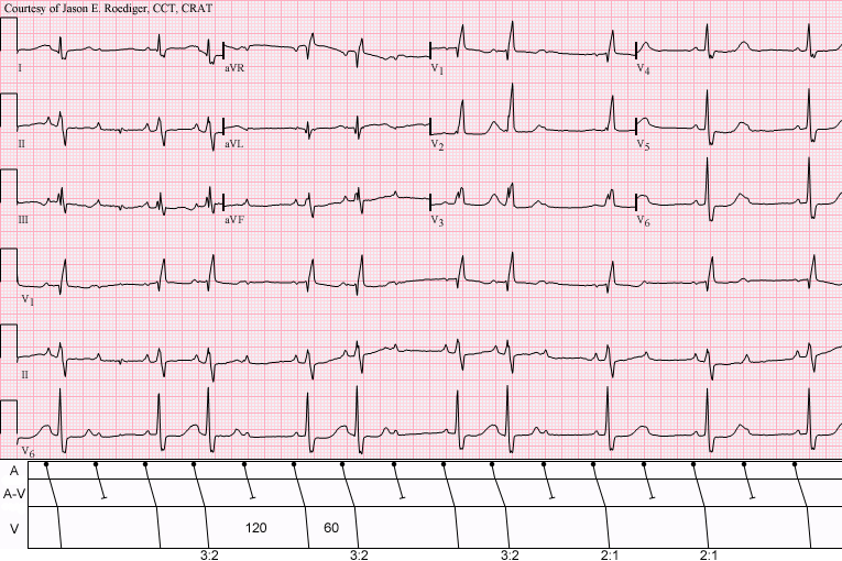
What does this tracing show? Choose the correct answer from the list below.
(1.) Sinus bradycardia with atrial bigeminy; conducted APBs; prominent U-waves; RBBB
(2.) Sinus rhythm with 3:2 and 2:1 Type II AV block; RBBB
(3.) Sinus rhythm with atrial bigeminy; both conducted and nonconducted APBs; RBBB
Acronyms:
APBs = atrial premature beats
RBBB = right bundle-branch block
Rate this content:
-

- jer5150's blog
- Log in or register to post comments
All our content is FREE & COPYRIGHT FREE for non-commercial use
Please be courteous and leave any watermark or author attribution on content you reproduce.
Comments
Number 2, I would say.
Number 2, I would say.
zafer karabulut
I think it's #3. The rSR' in
I think it's #3. The rSR' in V1 gives us the RBBB. The last 3 complexes show the Sinus Bradycardia. The first part of the tracing shows APBs, some of which are conducted, some of which are not. The 2nd last complex has an early, non-conducted p-wave burried in the t-wave (you can just see the little notch in the V6 rhythm strip).
Sarah
Mobitz II (Choice 2)
Atrial activity is best seen in lead II. The P waves overall look to be regular - ergo this is neither atrial bigeminy nor PACs. Instead we see groups of beats - but there ARE consecutively conducted complexes on occasion (3:2 cycles) with a constant PR interval - ergo Mobitz II.
Nice tracing!
Ken Grauer, MD www.kg-ekgpress.com [email protected]
INTERPRETATION
The correct answer is "2".
INTERPRETATION:
(1.) Sinus rhythm (rate about 100/min) with . . .
(2.) . . . 3:2 and 2:1 Type II A-V block.
(3.) Right bundle-branch block (RBBB).
COMMENTS:
Note that since the sinus rate is regular, the longer ventricular cycles (120) are exactly twice that of the shorter ventricular cycles (60). Conversely, in Type I A-V block, you would expect the longer ventricular cycles to be less than twice the shorter ventricular cycles. In the latter half of the tracing, during the 2:1 A-V block, one would strongly suspect Type II A-V block because the conducted beats display a normal P-R interval and RBBB is present but what clinches and proves that this is Type II A-V block are the consecutively conducted beats with unchanging P-R intervals.
This ECG represents both a bigeminal rhythm (i.e., 3:2 A-V block) and a regular bradycardia (i.e., 2:1 A-V block).
Type II A-V block is defined as the following:
CONSECUTIVE atrial impulses are conducted with identical P-R intervals before the "dropped" beat.
Jason E. Roediger - Certified Cardiographic Technician (CCT)
[email protected]