
Submitted by Dr A Röschl on Wed, 05/31/2023 - 02:02
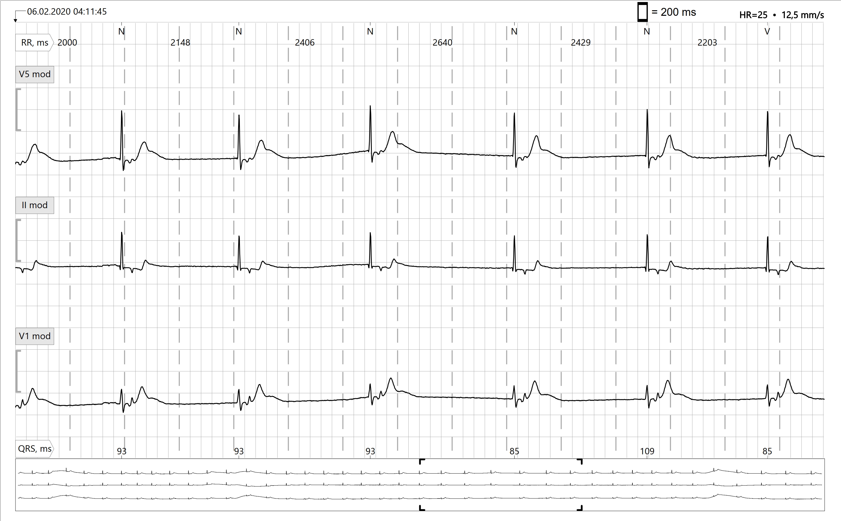
This ECG comes from a 75 yo man who had 2 syncopes in the past few weeks. The 12-lead-EKG at the family doctor showed an inconspicuous finding. Here you can see a section of the patients Holter ECG. There is a very slow junctional escape rhythm. How can this be recognized?
In sinus rhythm, there is a P wave in front of each QRS complex. The sinus P wave in the Holter-ECG is mostly concordant in at least 2 out of 3 leads (exeption: extreme heart axis), which means it goes in the same direction as the associated QRS complex. In V1 mod. the sinus P wave can be concordant or discordant.
In this EKG, the P wave is not before, but after the QRS complex, and it shows in the upper two leads as discordant, or opposite, the QRS complex. Depending on how deep the junctional center is located or how long the electrical impulse from this center needs to travel up to the atria and down to the ventricles, the retrograde P wave can be located before the QRS, hidden in the QRS or after the QRS. The latter is the case here. The low heart rate of 25 bpm is unusual for a junctional rhythm. A normal junctional rhythm has a heart rate between 40 and 60 bpm. In view of the history and the proven ECG changes, you have to assume that the patient quickly needs a pacemaker.
Rate this content:
-

- Dr A Röschl's blog
- Log in or register to post comments
All our content is FREE & COPYRIGHT FREE for non-commercial use
Please be courteous and leave any watermark or author attribution on content you reproduce.