
Submitted by Dr A Röschl on Sat, 11/15/2025 - 05:40
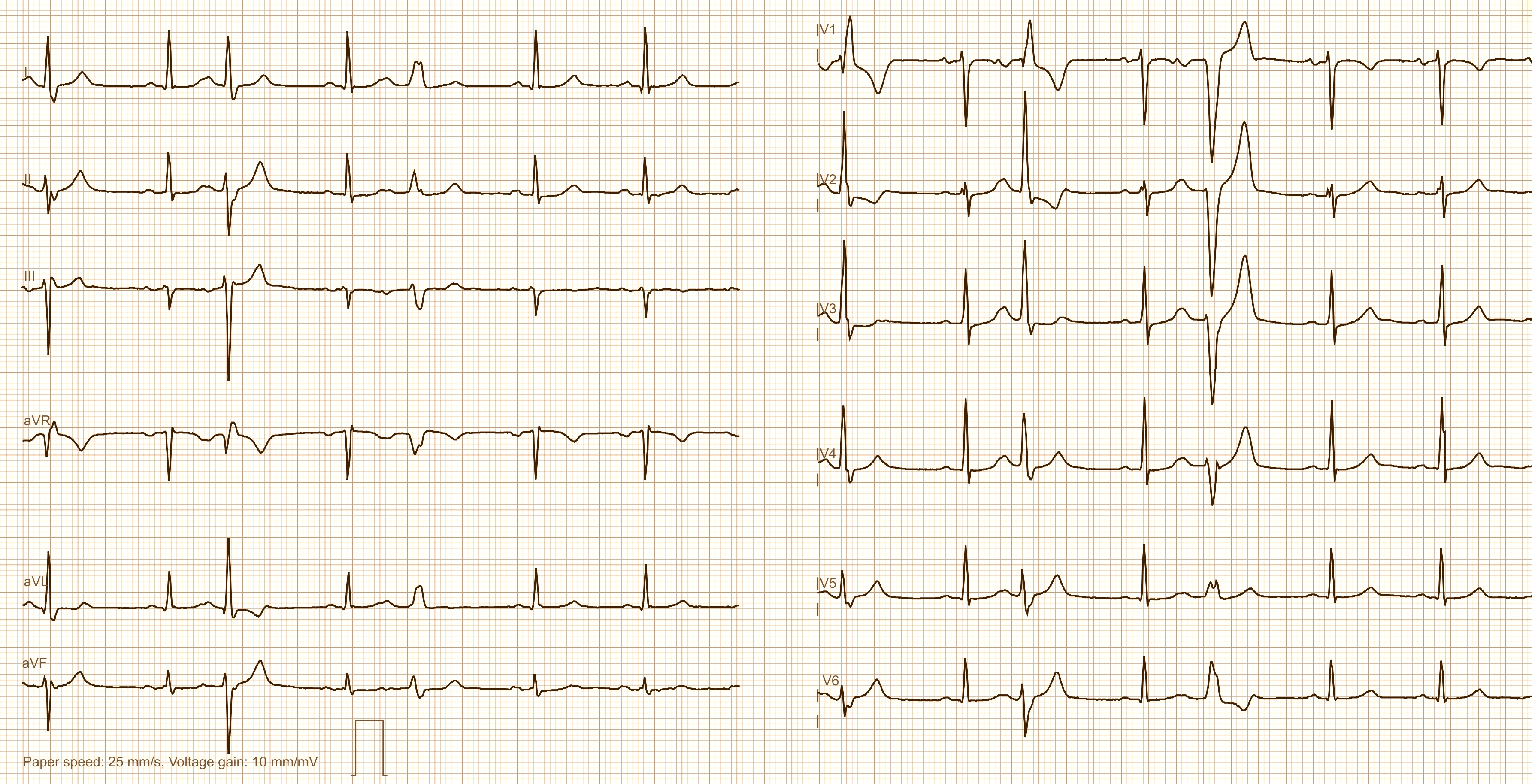
When wide QRS complexes appear in an ECG, the question always arises as to whether they are supraventricular or ventricular in origin. The distinction may not be so important in the case of individual extrasystoles, but it is certainly important in the case of tachycardia. Ventricular tachycardia is always a serious finding, whereas SVT with aberrant conduction is initially considered harmless.
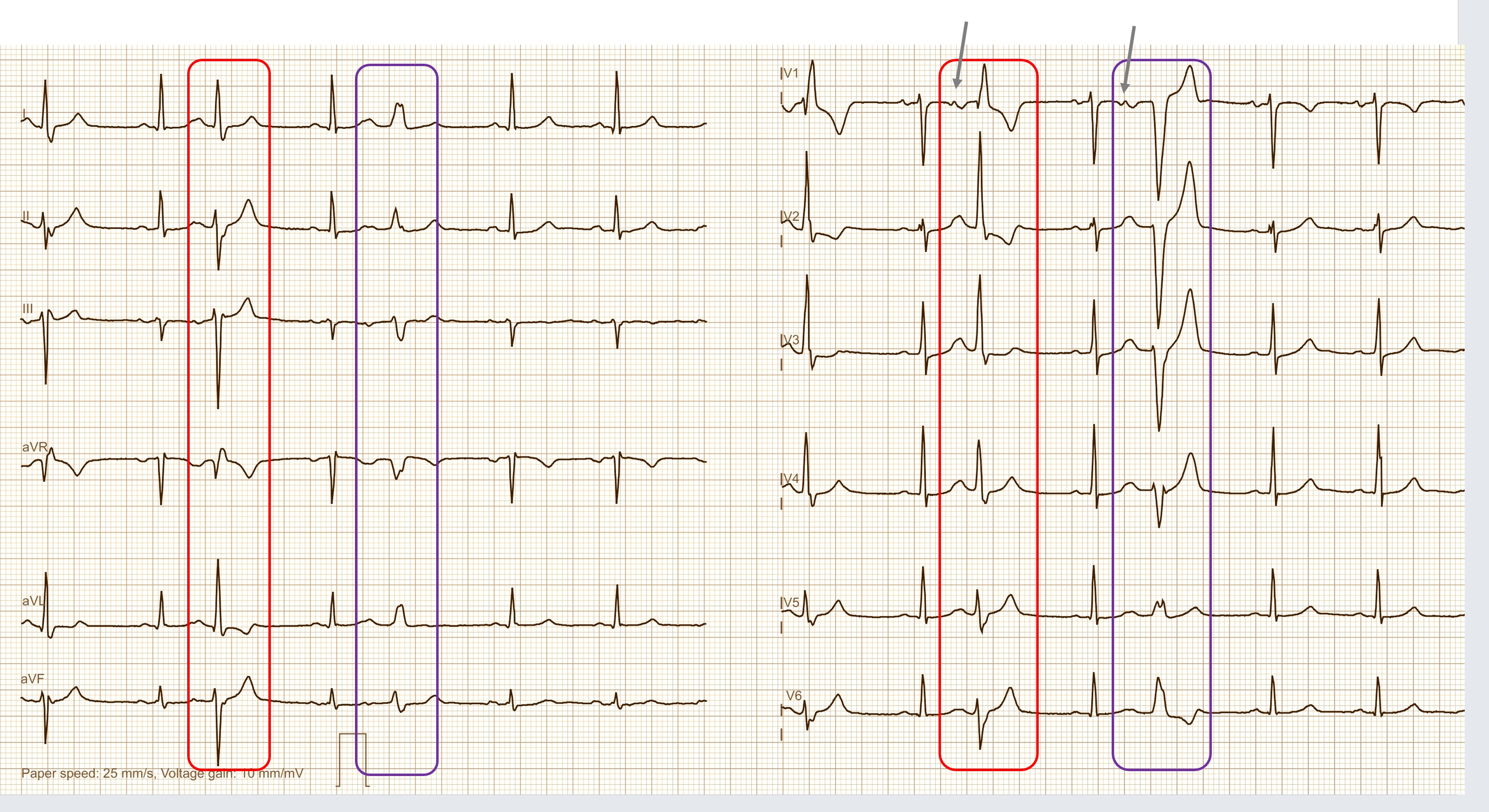
This example shows a sinus rhythm with several wide QRS complexes. The beat marked in red shows the typical morphology of an RBBB+LAFB, while the one marked in blue shows that of an LBBB.
In V1, the corresponding premature P wave is very easy to recognize, indicating PACs with aberrant conduction rather than PVCs.
The first QRS complex is not marked, but shows a similar picture to QRS number 3. Here, too, there is a QRS complex with the pattern of a RBBB and LAFB, but with a slightly different QRS morphology. We cannot see the P wave preceding the first QRS complex, but we can assume that it is present and that there is also a PAC with aberrant conduction here.
Rate this content:
-

- Dr A Röschl's blog
- Log in or register to post comments
All our content is FREE & COPYRIGHT FREE for non-commercial use
Please be courteous and leave any watermark or author attribution on content you reproduce.