
Submitted by Dawn on Sun, 08/23/2015 - 12:20
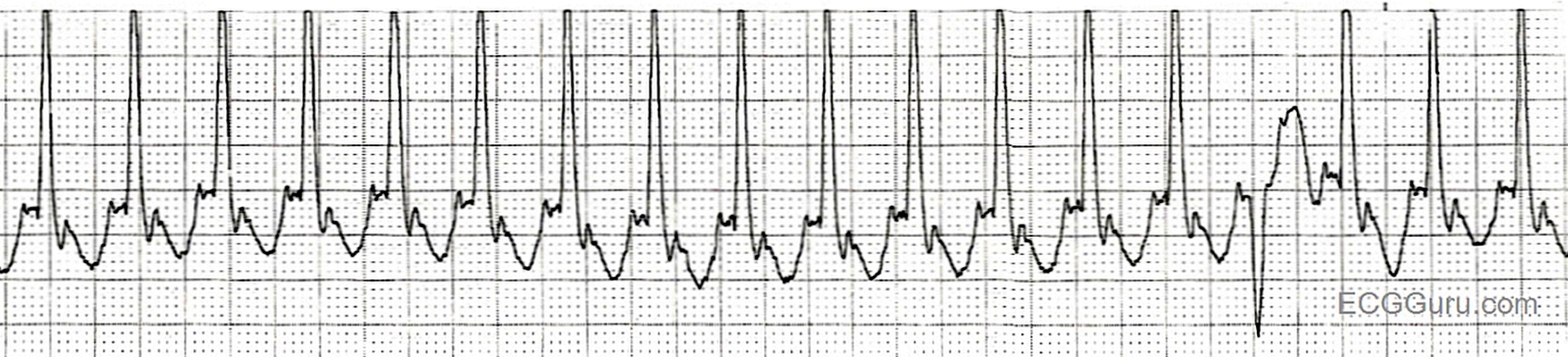
This strip was taken from a patient at rest. It shows a regular tachycardia with a slightly-widened QRS complex at about .10 seconds duration. It is somewhat difficult to evaluate the baseline for P waves or flutter waves. We ALWAYS recommend multi-lead assessment for such evaluation. The P waves (or flutter waves) here have a sharp point, and can be easily "marched out", with a rate of about 300 per minute.
Whenever the ventricular rate is near 150/min., we should always consider the possibility of atrial flutter with 2:1 conduction. Since atrial flutter results in atrial depolarization at around 250 - 350 per minute, conducting every other P wave results in a rate of about 150. It can masquerade as sinus tach, but a patient with sinus tach at such a fast rate would probably have an obvious cause for a rapid heart rate, such as hypovolemia, drug overdose, or exertion. This rhythm could also be mistaken for atrial tachycardia or other forms of supraventricular tachycardia (SVT, PSVT, AVNRT, etc.). Multiple leads can more easily uncover the flutter waves running continuously "behind" and "through" the QRS complexes.
There is one beat that is obviously different from the others. This beat is about the same width as the other QRS complexes, but is opposite in direction. This probably represents aberrant conduction, possibly a hemiblock that occurs only in this beat. Careful measurement will show that this QRS is very slightly early, while the others are all very regular. The slight width of all the QRS complexes suggests that there is a conduction delay, which cannot be diagnosed on one strip with no patient history available.
There are other differential diagnoses, such as ventricular tachycardia with a captured sinus beat. We welcome discussion of this interesting strip.
Related Terms:
Rate this content:
All our content is FREE & COPYRIGHT FREE for non-commercial use
Please be courteous and leave any watermark or author attribution on content you reproduce.
Comments
fusion beat?
This is quite a trivial point, but I think that the ‘different’ QRS is a ventricular premature, or more likely, fusion beat. It could be, as Dawn says, an example of intermittent aberrant conduction, but this usually happens for a reason, eg a change in atrial rate. The QRS following the different one appears very slightly delayed, consistent with retrograde concealed conduction of a ventricular impulse into the AV node. The reason the different QRS is not abnormally broad is probably because it is actually a fusion beat. This is a bit conjectural, though, because it’s probably not possible to be sure of the origin of one beat in a single-lead rhythm strip. The important thing here is to recognise that the rhythm, as Dawn points out, is atrial flutter.
Dave R
AFlutter with a Different-Looking Beat (Aberrant vs PVC?)
Ken Grauer, MD www.kg-ekgpress.com [email protected]