Displaying 21 - 26 of 26
Dawn's picture

Third-degree AV Block and Junctional Escape Rhythm With Right Bundle Branch Block and Prolonged QTc Interval

This ECG is from a 70 year old woman for which we have, unfortunately, no clinical information.  It shows a sinus rhythm with a rate of about 72 bpm (NSR) with AV dissociation caused by third-degree heart block.  The escape rhythm is junctional at a rate of 38 bpm.  There appears to be a right bundle branch block, based on the QRS duration of 132 ms, and a wide S wave in Leads I and V6.  The precordial leads do not show the usual RBBB pattern of rSR' in V1 and V2, and the r wave progression is poor (non-existent).  This is felt to be due to poor lead placement (a good teaching point).  Of interest, the ECG machine has reported a "severe right axis deviation" based on the tall upright R wave in aVR and the deep S in avF.  In RBBB, the first part of the QRS represents left ventricular depolarization, and the terminal wave represents the delayed right ventricle.  In effect, the two ventricles have their own electrical axes, which we can see because the ventricles are not depolarizing simultaneously.  The axis of the LV appears to be normal in this tracing.

In addition to the above, this patient has a very prolonged QT interval.  The QT is longer in bradycardic rhythms, but when corrected to a standard of 60 bpm (QTc), this patient's QT interval is still prolonged at QTc: 552 ms.  Without clinical data, we cannot speculate  as to why this patient's QTc is prolonged, but it can be a very dangerous situation.  Follow the links for more information on QT prolongation and Torsades de Pointes and Long QT Syndrome.

As always, we welcome comments from our members adding insight to this interesting ECG, and also questions you would like to ask our Guru members.

Dawn's picture

Right Bundle Branch Block and Previous Anterior Wall M.I.

This example of previous anterior wall M.I. with right bundle branch block is from a 95-year-old man.  He survived an anterior-septal M.I. caused by an occlusion of the left anterior descending branch of the LCA. The septum was involved in the damaged area, and the patient was left with a right bundle branch block.  The pathological Q waves in Leads V1 through V3 show the location of permanent damage, or necrosis.

In this case, the usual rSR' pattern of right bundle branch block is replaced by qR, indicating loss of the initial r wave due to the M.I.

Review of RBBB criteria:  wide QRS, rSR' pattern in V1, and Rs with a slurred s wave in I and V6.  These criteria must be present in a SUPRAVENTRICULAR rhythm to diagnose RBBB.

Dawn's picture

Right Bundle Branch Block

This ECG is from a healthy young man in his 20's.  He was born with a ventricular septal defect (VSD) that was surgically repaired when he was a toddler.  He now has a right bundle branch block, which could be a result of the defect, or the surgery.  This is a good ECG for the Instructors' Collection because it clearly shows all the ECG characteristics of right bundle branch block:  wide QRS in a supraventricular rhythm (in this case, NSR), rSR' pattern in V1, wide or "slurred" S waves in Leads I and V6.  There is no rhythm strip below the 12 leads in this ECG, but there is no rhythm disturbance.

Dawn's picture

Left Bundle Branch Block In Patient With Severe Aortic Stenosis

This ECG is from a 91-year-old man who was being evaluated for replacement of his aortic valve, which was severely calcified. It shows a classic LBBB pattern: wide QRS, supraventricular rhythm (normal sinus rhythm with first-degree AV block), a negative QRS in V1, and a positive QRS in Leads I and V6.

Dawn's picture

Intermittent Left Bundle Branch Block

This patient suffered a recent anterior-septal wall M.I., which can be seen as slight ST elevation in V1 and ischemic T wave inversions in V2 through V4. The patient has developed an intermittent left bundle branch block as a result of this M.I.  Every other beat is conducted in a left bundle branch block pattern, as the LBB cannot repolarize in time for each beat.  The criteria for LBBB are:  wide QRS, supraventricular rhythm, and negatively-deflected QRS in V1 with a positive QRS in V6 and Lead I.

Dawn's picture

Atrial Flutter With 1:1 Conduction and Rate-dependent Right Bundle Branch Block

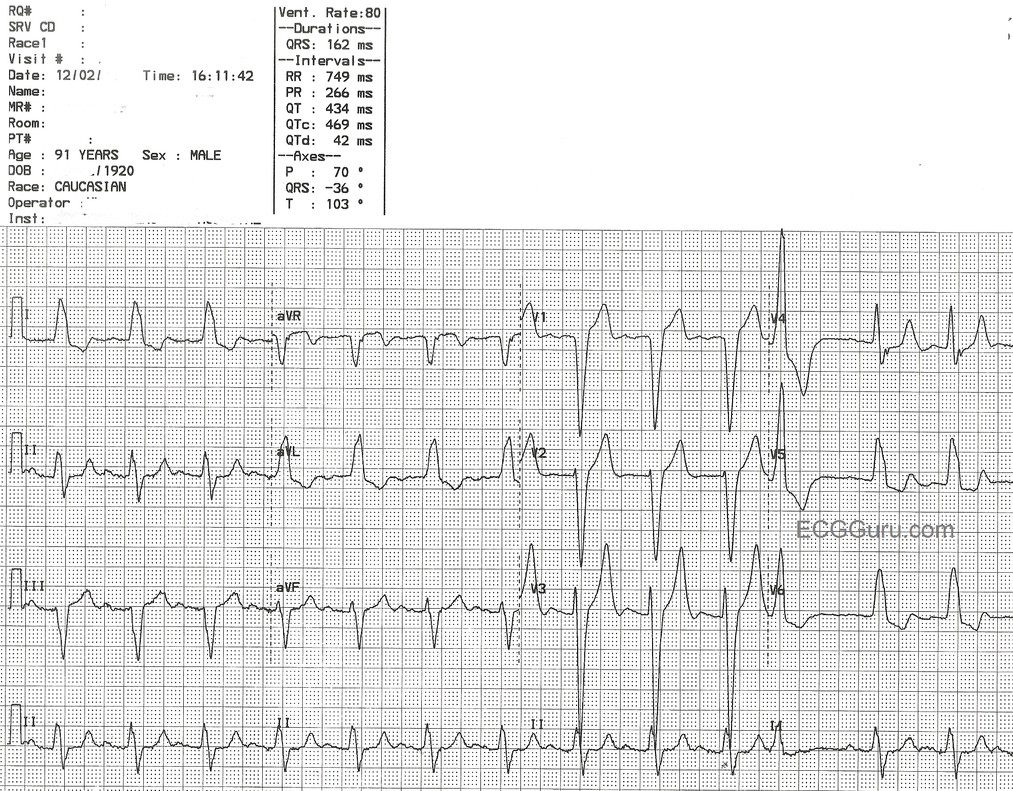
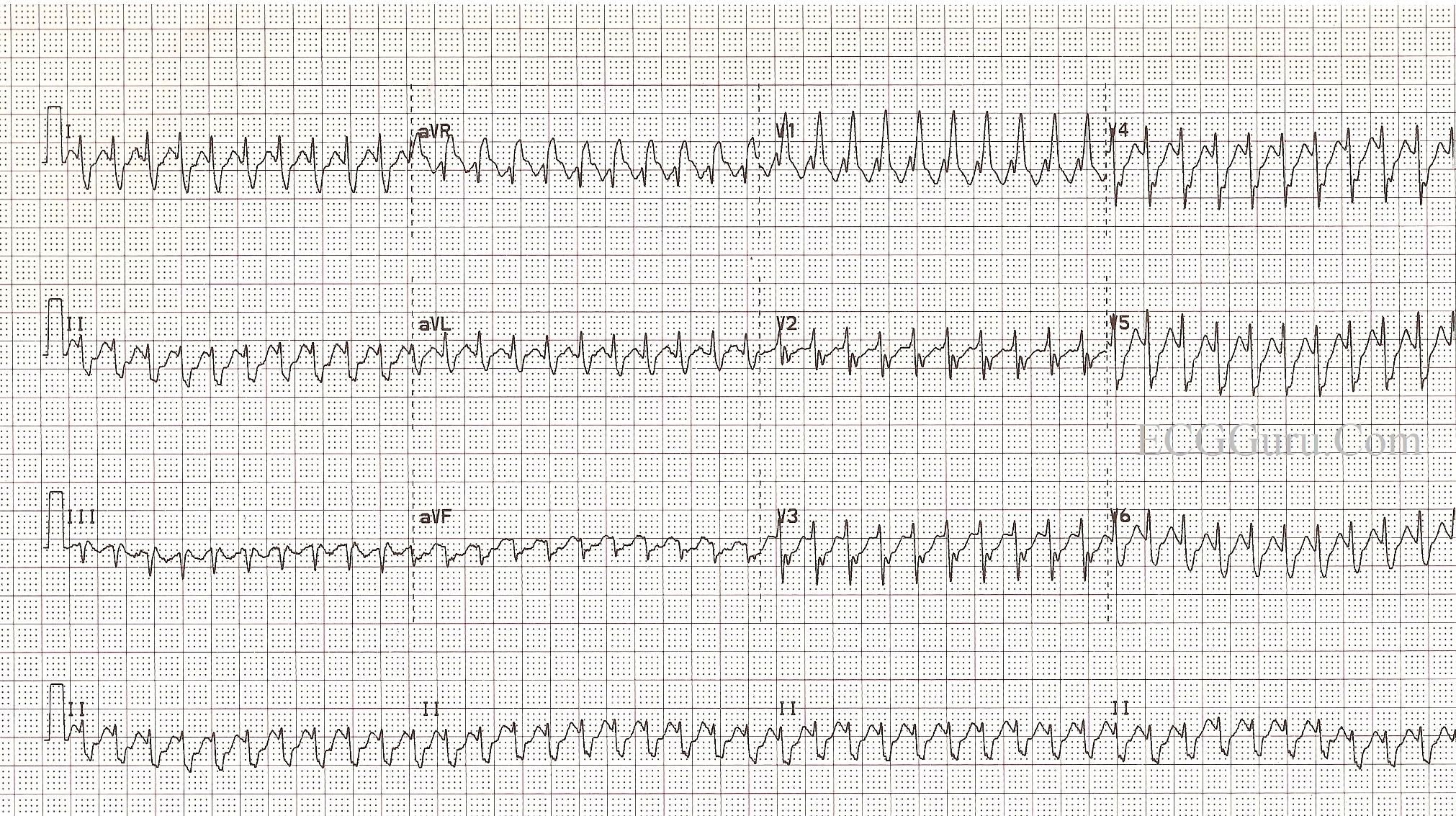
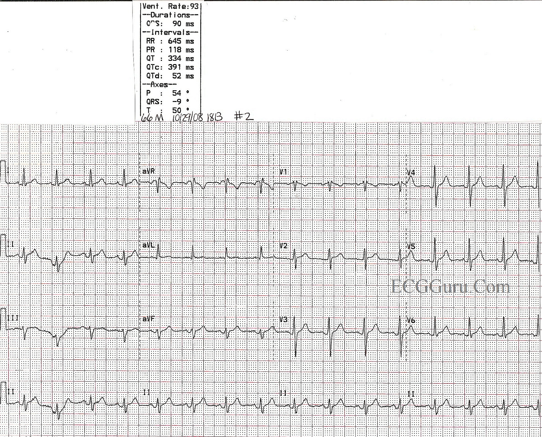
The first ECG is from an active, otherwise healthy 66-year-old man who experienced a sudden onset of symptomatic tachycardia. He presented to the Emergency Dept. feeling (understandably) very anxious, with poor perfusion to his skin, chest discomfort, and palpitations. As the ED staff prepared to electrically cardiovert him, he spontaneously converted to normal sinus rhythm at a rate of 93 / min. and a QRS duration of 90 ms.

After a normal diagnostic cardiac cath, he was sent to the EP lab and the circuit responsible for this rhythm was successfully ablated. He was discharged in good condition the next day.

This is a good ECG for all levels of students. For beginners, it shows that atrial flutter is one of the rhythms we call "SVT", and it does not always conduct in a variable rate, or a rate that allows "sawtooth" P waves to show easily. You can show them that the "sawtooth" pattern, brought about by P waves that are 250 - 350 per minute, is still there if one ignores the QRS complexes for a moment. This is a great ECG for discussions of rate and cardiac output, and making decisions regarding treatment based on the patient's hemodynamic condition. For more advanced students, this ECG can lead to a discussion of the accessory pathways and re-entrant pathways that cause rapid rhythms. For RBBB criteria, click HERE.

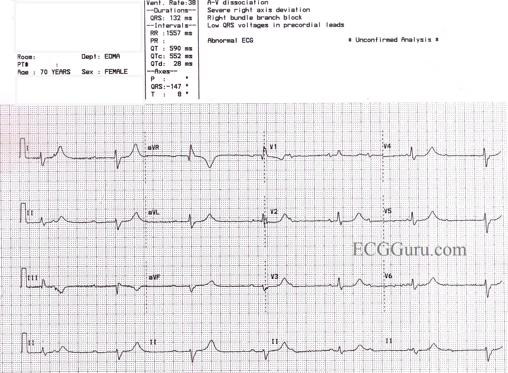
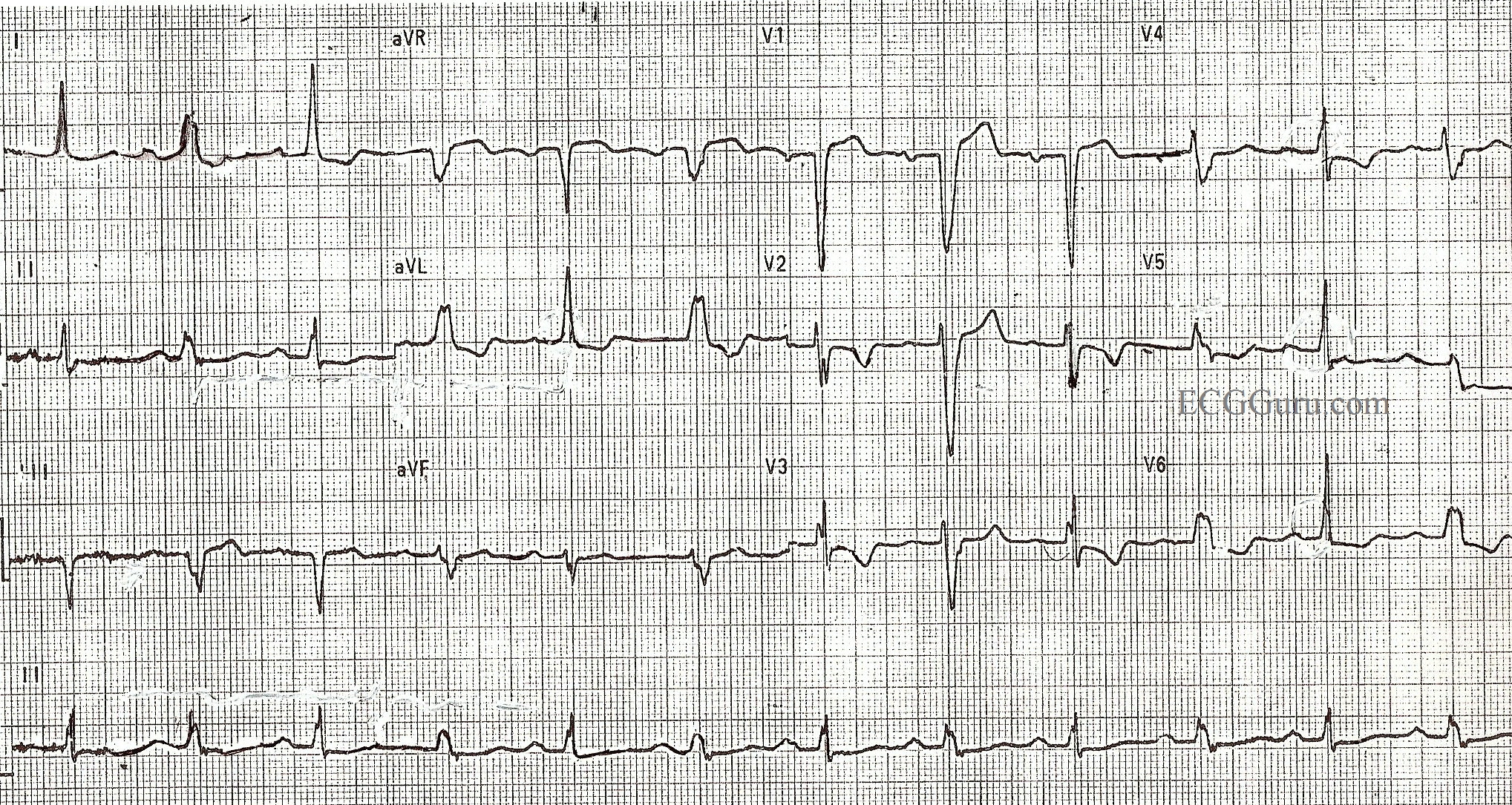
The second ECG shows the same patient after spontaneous conversion to sinus rhythm.  The bundle branch block has disappeared, as it was rate-dependent.

Pages

All our content is FREE & COPYRIGHT FREE for non-commercial use

Please be courteous and leave any watermark or author attribution on content you reproduce.