Dawn's picture

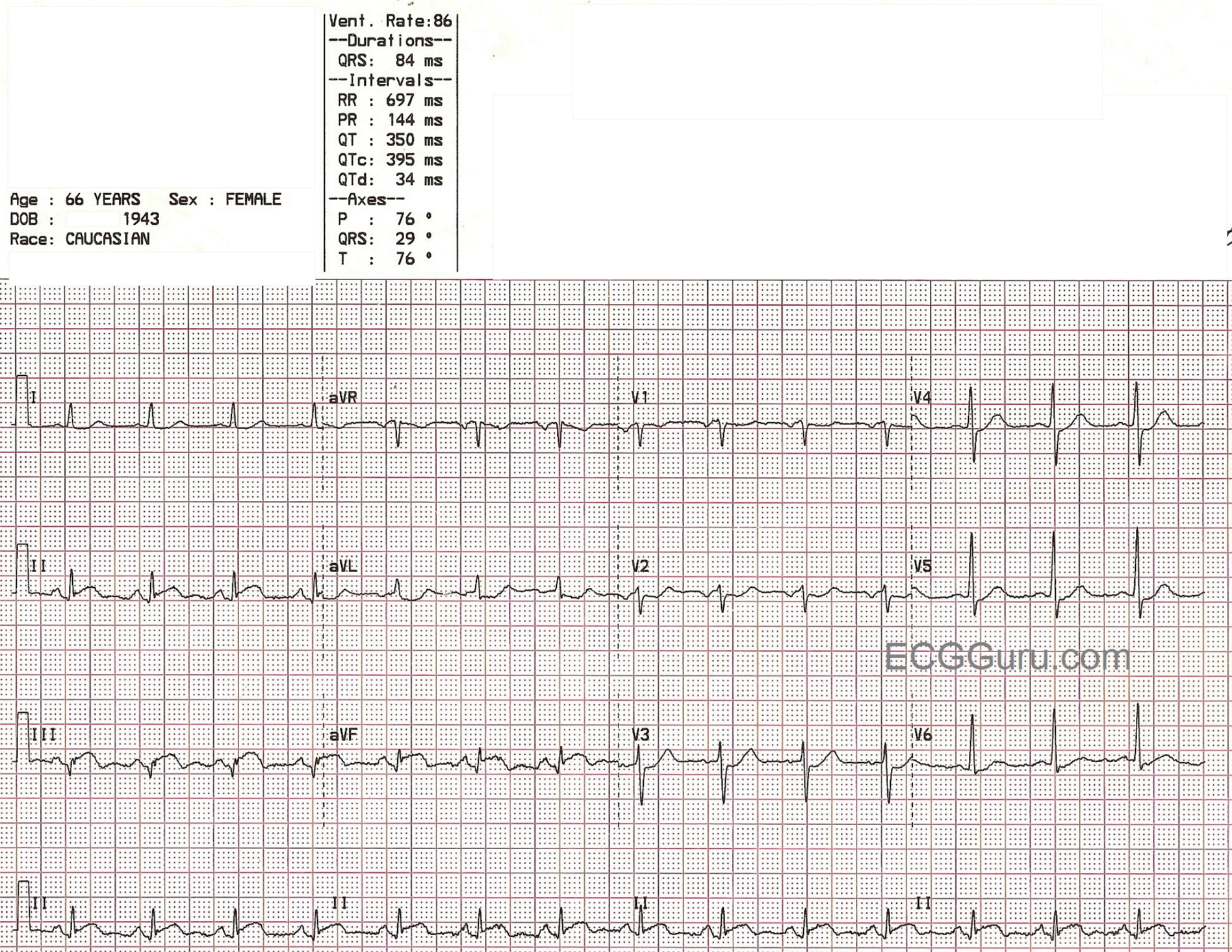
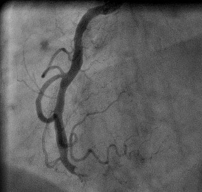
This ECG was taken from a 66-year-old woman who presented to the emergency department complaining of chest pain and shortness of breath.  She attributed her symptoms to her COPD, but stated that her aerosol treatment had not helped.  She waited for some time before deciding to go to the hospital, then drove herself. In the emergency department, she had blood drawn, an I.V. started, and an aerosol treatment of albuterol.  She was then taken to the radiology dept. for a chest xray.  When she returned, a 12-lead ECG was done.  45 minutes had passed since she first arrived at the hospital.  This ECG is shown here.  It shows ST elevation in the inferior leads:  II, III, and aVF.  The patient was taken immediately to the cath lab, where her right coronary artery was found to have a 100% occlusion. (See image accompanying this ECG).  Angioplasty was successful, and stents were placed in the artery.  The patient was found to have anemia, with a hemoglobin of 5.5, and she was given a blood transfusion.  During the cath procedure, a ventriculogram was performed, which showed a stunned and akinetic inferior wall.  Unfortunately, subsequent ventriculogram performed several weeks later showed the lack of motion of the inferior wall to be permanent.  The ventriculogram is also posted here, and at this YouTube link, so you will be able to show your students the mechanical effects on the heart of a delay to treatment in acute STEMI.  See our YouTube site for more ventriculograms and cath videos.

Rate this content: 
Average: 3.5 (4 votes)

Comments

ekgpress@mac.com's picture


There are a number of important messages integrated in Dawn's Instructor ECG for this Week:

  • A highly illustrative teaching ECG diagnostic of acute infero-postero MI. Inferior leads show marked coved ST elevation with beginning T wave inversion in leads III and aVF. Q waves have already formed in all 3 inferior leads. There is reciprocal ST depression - which is subtle in a number of leads, and most marked in lead aVL. Proximal RCA occlusion is strongly suggested by ST elevation in III > II - and - marked ST depression in aVL > I. There is also anterior ST depression of shape consistent with a positive "mirror test" - which suggests associated posterior wall involvement. There is no suggestion on this tracing of RV involvement (no subtle ST elevation in lead V1) - though right-sided leads would be needed to better assess this if indicated.
  • The fact that substantial Q waves are already present in the inferior leads of this tracing suggests that some time has already passed since onset of the infarct ...
  • This 66-year old woman had onset of chest pain and shortness of breath - but failed to realize she was having a heart attack for some time. Such delay is still all too common among too many individuals ...
  • This patient also had significant anemia (Hgb = 5.5) that she was also unaware of - and which doubtlessly contributed (if not caused) her event. A slow GI bleed is often relatively asymptomatic - even to very low hemoglobin levels IF the rate of bleeding is slow. Important for health care providers to always consider the possibility of anemia whenever an older patient presents with chest discomfort, weakness or shortness of breath.
  • Even if you are inexperienced in viewing cath films - the examples Dawn posts here of a normal angiogram - and of this patient's angiogram with loss of inferior wall motion are unmistakable. Please check out both examples that Dawn posts here (total time = less than 15 seconds to view).
  • Had this patient not delayed so long - had she not attributed her symptoms to her COPD and had she realized she was increasingly anemic (most such patients retrospectively acknowledge weakness and dizziness that they had not realized were due to anemia) - much myocardium could have been saved.
  • For more on Infarct Localization - CLICK HERE
  • RIGHT-CLICK HERE to download more on the "MIRROR TEST".

Ken Grauer, MD  www.kg-ekgpress.com   [email protected] 

All our content is FREE & COPYRIGHT FREE for non-commercial use

Please be courteous and leave any watermark or author attribution on content you reproduce.