Displaying 11 - 20 of 33
Dawn's picture

Left Bundle Branch Block With Second-Degree AV Block, Type II

 This ECG was obtained from an 84-year-old woman who was scheduled for surgery.  When the anesthesiologist did this ECG, the surgery was cancelled. It is a very good example of fascicular-level blocks. 

The underlying rhythm is a regular sinus rhythm at about 95 bpm.  There are some non-conducted P waves which are part of the sinus rhythm (not premature beats).  When the P waves DO conduct, the PR interval is steady at about .15 seconds (148 ms).

In addition, there is a LEFT BUNDLE BRANCH BLOCK.  The ECG criteria for LBBB are:  1) A supraventricular rhythm, 2) A wide QRS, and 3) A negative QRS in Lead V1 and a positive QRS in Leads I and V6.  The QRS duration in this ECG is 136 ms.

There are generally two fascicles (branches) in the left bundle branch, and one main fascicle in the right bundle branch.  So, a LBBB represents a “bi-fascicular block”.  That means that A-V conduction is proceeding down only one fascicle (the right bundle branch).  In that fascicle, there is an “intermittent” block.  When the RBB is not blocked, we see a QRS.  When it is blocked, we see none.  This is then termed an “intermittent tri-fascicular block” – otherwise known as SECOND-DEGREE AV BLOCK, TYPE II.  Type II blocks nearly always have a wide QRS due to the underlying bundle branch pathology.  You may see RBBB, LBBB, or RBBB with left anterior fascicular block (hemiblock).  Very rarely, the combination might include left posterior hemiblock.  The intermittent block in the “healthiest” fascicle(s) is what makes this a second-degree block, and not a complete heart block (third-degree AVB).

The clinical implications of this block are that the heart is operating on only one fascicle, and that fascicle is showing obvious signs of distress.  A third-degree AVB could be imminent.  In addition, LBBB causes a wide QRS, which decreases cardiac output.  Second-degree, Type II AVBs can result in very slow rates, and sometimes cause more hemodynamic instability that some third-degree AV blocks.

This patient was scheduled for pacemaker implantation instead of the originally-scheduled surgery. 

Dawn's picture

AV Block With Changing PR Intervals

Just like other subjects we are taught in school, ECG interpretation is usually taught in a very basic, simplistic way.  As we add to our knowledge, we are able to determine the mechanisms of more complex rhythms. 

When I took my first basic ECG rhythm monitoring course, I memorized all the “rules”, and at the end of the course, I thought I could read ANY strip correctly.  Then, in real life, I found that some rhythms can’t be interpreted from one lead, or even from one 12-lead ECG. 

This strip offers advanced readers to challenge themselves, and it offers teachers a chance to show students an “exception to the rules” if it is appropriate for those students.  We all learn the classification of second-degree AV blocks:  Both Type I and Type II show an underlying sinus rhythm with some P waves conducted and some not.  Type I has progressively prolonging PR intervals until a P wave is non-conducted.  The cycle restarts after the dropped QRS.  Type II has PR intervals that are all the same, and may be prolonged or normal. 

In this ECG, you will be able to “march out” a normal sinus rhythm at a rate of 80 bpm.  The P waves are marked with small dots at the bottom.  Two of every three P waves are followed by QRS complexes.  Is it Type I?  No – the PR intervals are not prolonging.  Is it Type II?  The PR intervals are not the same!  What is happening? 

There is also left bundle branch block, which is a sub-Hisian block.  Blocks occurring in the intraventricular conduction system include bundle branch blocks, second-degree AVB Type II,  and third-degree AVB with ventricular escape.  This group of blocks tends to be more threatening than the blocks that occur in the AV node (second-degree type I and third-degree with junctional escape). 

Dawn's picture

Left Bundle Branch Block With Left Ventricular Hypertrophy

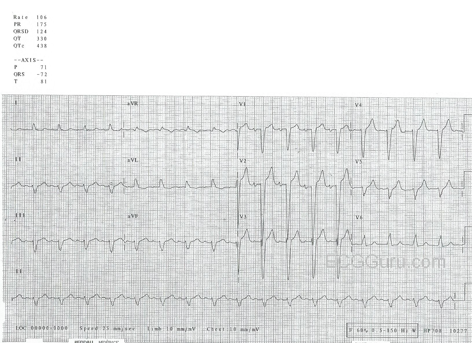
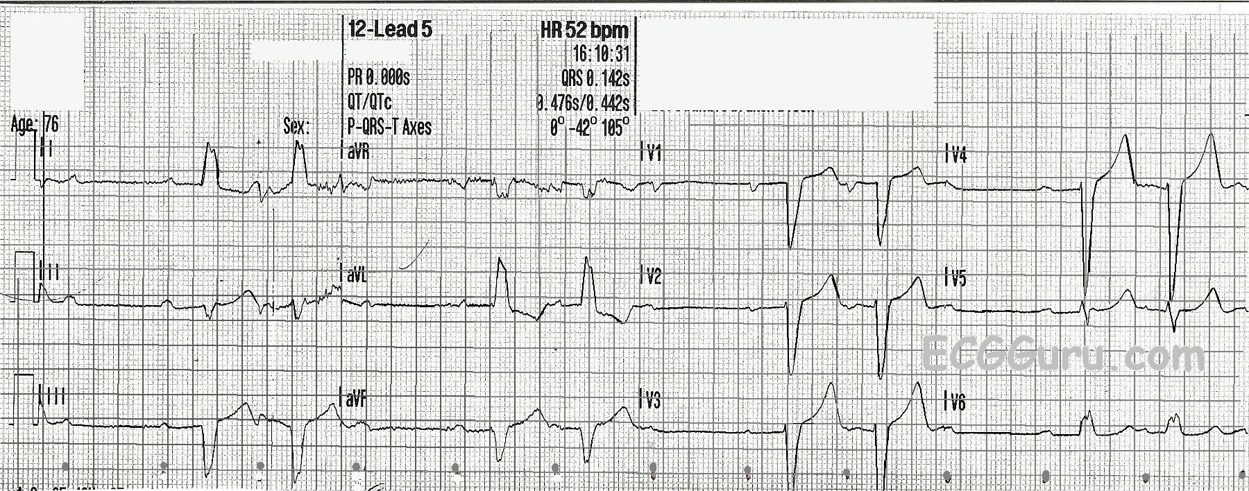
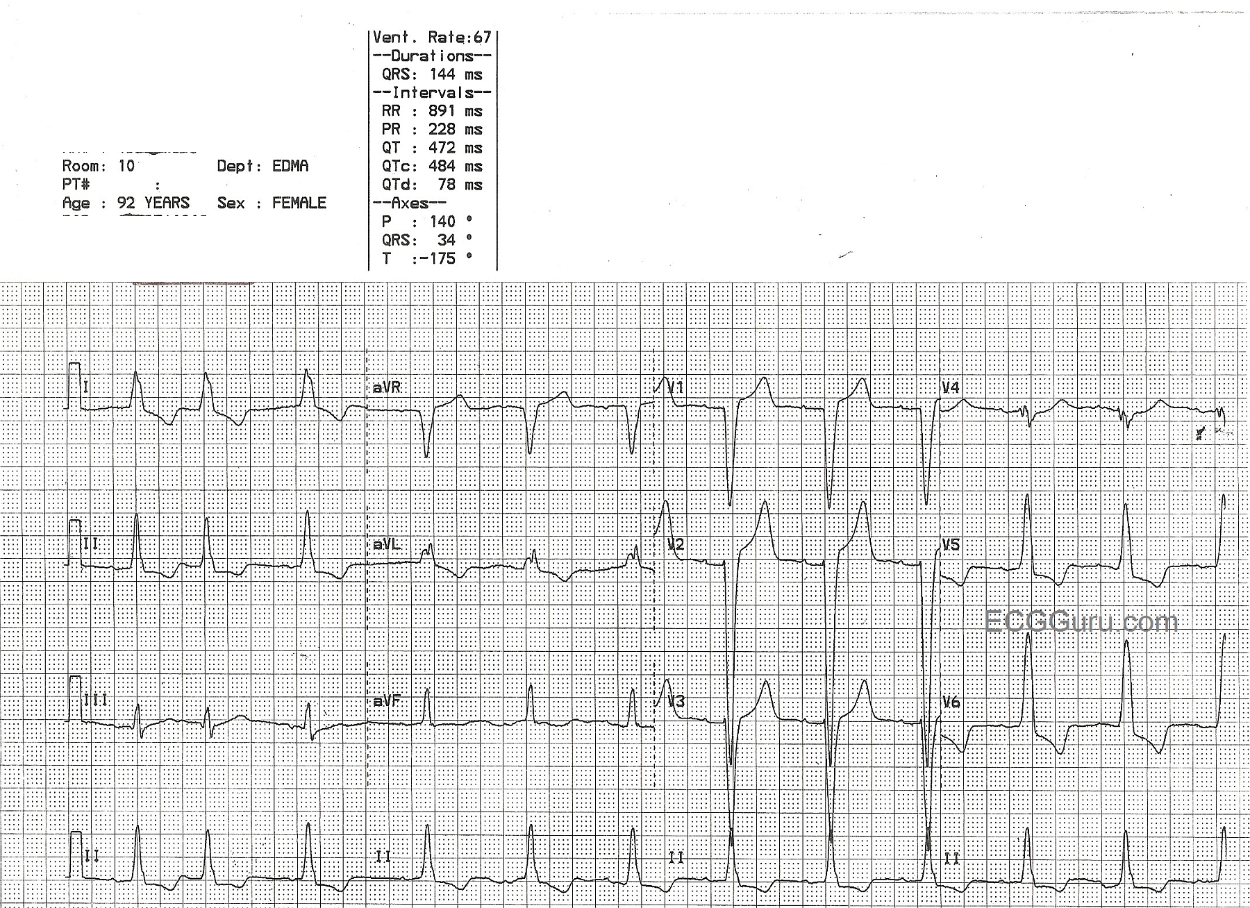
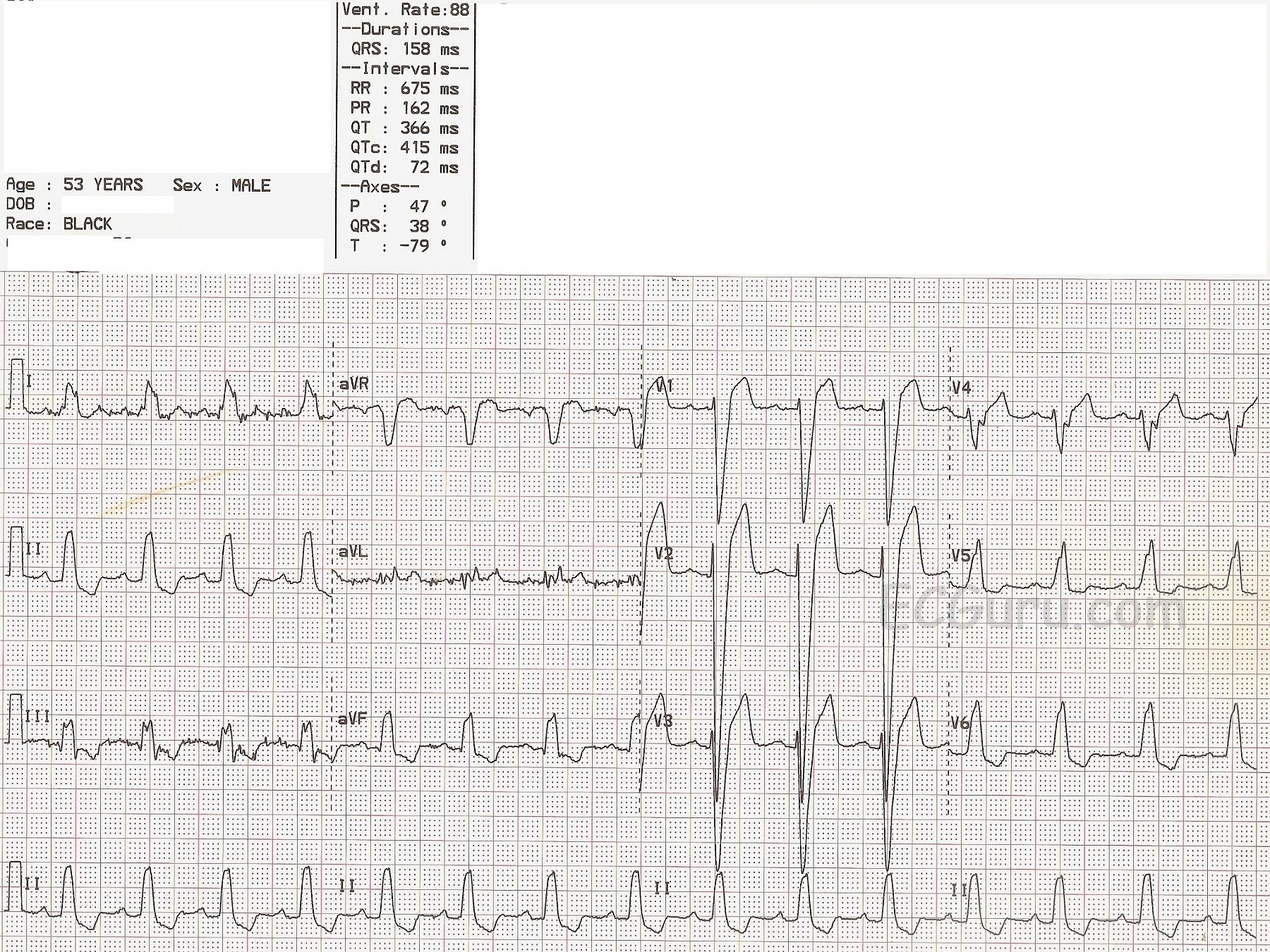
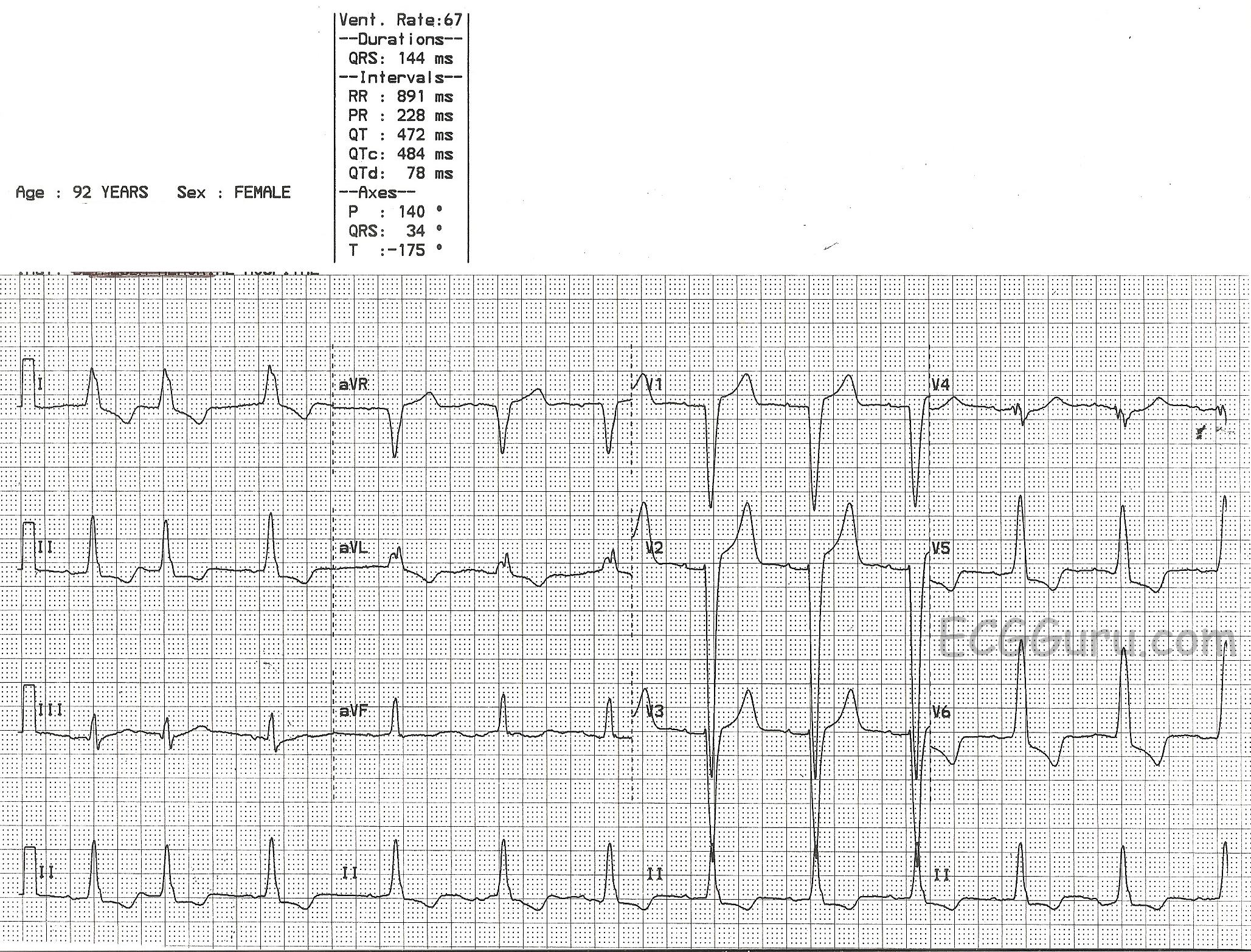
This 92-year-old patient was diagnosed with left bundle branch block on ECG, and left ventricular hypertrophy on echocardiogram.  The two conditions are very often seen together, in fact, a majority of LBBB patients have LVH.  Since the two conditions can have similar ECG changes, it is difficult from the ECG alone to determine the presence of LVH when LBBB is present.  If the ECG criteria for LVH are present, it can be assumed that LVH is present, even in the presence of LBBB.  For determining LVH by ECG criteria, the Sokolov-Lyon criteria are commonly used ( S wave in V1 + R wave in V5 or V6 > 35 mm). 

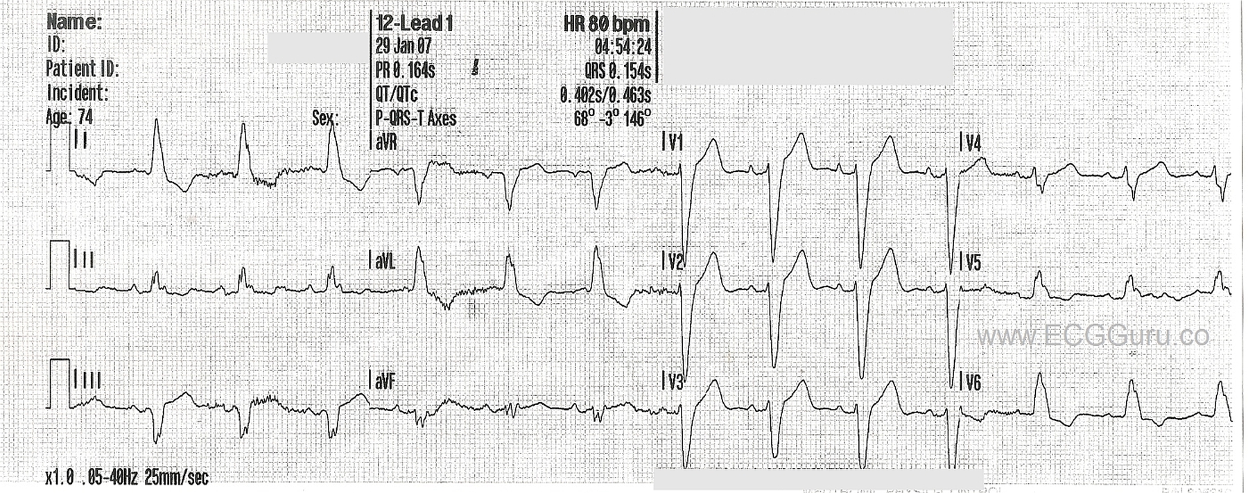
The common criteria for left bundle branch block include:  wide QRS complex, frontal plane axis normal or leftward, negative QRS in V1, and positive QRS in leads I and V6.   LBBB is only found in supraventricular rhythms (not ventricular rhythms).  The ST segment and T wave will be negative in leads with positive QRS complexes, and positive in leads with negative QRSs (discordant).

Left ventricular hypertrophy also widens the QRS, although not often as much as LBBB does.  There will be discordant ST segments and T waves, which is called the "strain" pattern.  It also is easier to diagnose in supraventricular rhythms, because ventricular rhythms usually have large QRS complexes due to the depolarization wave being in one direction across the heart.

For confirmation of LVH, an echocardiogram is recommended.

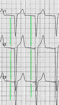
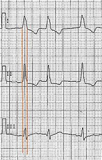
This ECG also has an interesting rhythm.  The first beat appears normal, the second beat is a PAC.  The third beat appears to arise from a different focus, which would make it an escape beat, but it is very difficult to determine this due to the very tiny P waves.  After a pause, a regular sinus rhythm resumes.  To see the P waves, look at the right chest leads:  V1, V2, V3.  Since left bundle branch block only occurs in SUPRAVENTRICULAR rhythms, it is important to determine the rhythm, and P waves are a definite sign of SV rhythm.  We wish the P waves here were taller.

Dawn's picture

Atrial Fib To Cardiac Arrest

A paramedic crew responded to the office of a local physician. A 61-year-old male presented with a one-week history of chest pain and shortness of breath. He had a previously undiagnosed atrial fibrillation with rapid ventricular response and left bundle branch block, but was alert. Shortly after transport commenced, the patient became unresponsive with Torsades de Pointes, which rapidly degenerated into ventricular fibrillation. The paramedic placed pads and defibrillated within one minute.  After two minutes of compressions, the patient had a fairly regular rhythm with return of spontaneous circulation.  Transport time was short.  On catheterization, the patient was found to have severe coronary artery disease, requiring coronary artery bypass graft surgery (CABG) A balloon pump was inserted in an attempt to strengthen him for surgery.

What is the rhythm?   The 12-lead ECG presented here shows atrial fibrillation at a rate of 138 per minute.  The rhythm is irregularly-irregular with no P waves.  Since the patient had not yet been diagnosed with atrial fib, obviously no therapy had been initiated to control the rate. There is a PVC near the end of the strip.

Dawn's picture

Left Bundle Branch Block

This ECG shows a “classic” left bundle branch block pattern. 

The ECG criteria for left bundle branch block are:

·        Wide QRS (.12 seconds or greater)

·        Supraventricular rhythm (ventricular rhythms do not travel via the LBB)

·        The QRS in V1 is negative, and the QRS in Leads I and V6 are positive. 

The left bundle branch (LBB) can be blocked permanently, temporarily, intermittently, or in the because of a fast rate.  When the LBB is blocked, conduction proceeds from the AV junction down the right bundle branch, depolarizing the right ventricle.  The impulse travels from the right ventricle across the left ventricle, cell by cell.  Conduction is slower this way, and there is asynchrony of the ventricles. This slow conduction and asynchrony of the two ventricles causes widening of the QRS complex.

NOTE:  It is "normal" for wide-complex rhythms to have ST segment elevation in leads with negative QRS complexes and ST depression in leads with positive QRS complexes.  This can make it a bit difficult to determine the ST changes of acute M.I. 


Dawn's picture

Left Bundle Branch Block and Artifact

This ECG offers several teaching opportunities.  First, it is an example of left bundle branch block (LBBB).  It was obtained from a 53-year-old man who was undergoing a cardiac cath for chest pain.  Unfortunately, we do not have access to his past medical history or the results of his cath.  The ECG criteria for a diagnosis of LBBB are:  1) wide QRS complex; 2) supraventricular rhythm; 3) negative QRS in V1 and positive QRS in V6 and Lead I.  This ECG shows normal sinus rhythm at a rate of 88 bpm and a wide QRS at 158 ms (.158 seconds).  The QRS in V1 is negatively deflected and in V6 and Lead I it is positive.

In LBBB, as with any condition that significantly widens the QRS, there will be ST-T changes.  The ST segment will deviate in the opposite direction of the QRS.  In other words, there will be ST elevation in leads with negative QRS complexes and ST depression in leads with positive QRS complexes.  LBBB causes significant difficulty for those trying to diagnose acute ST elevation using ECG alone.  Excessive ST elevation in a lead where elevation is expected OR ST elevation in a lead where depression is expected should be considered to be abnormal.  At this point, you may find it useful to review Sgarbossa's Criteria regarding determining the presence of acute M.I. in the presence of LBBB.

LBBB can be a serious functonal problem for the patient, as the slow ventricular conduction that causes the wide QRS results in less-than-optimal cardiac output.  This associates LBBB with congestive heart failure, both as a cause of CHF and a result of CHF.  Many people with LBBB and CHF can be helped by cardiac resynchronization therapy - pacing both ventricles synchronously to narrow the QRS and improve cardiac output.  For an excellent article on cardiac pacing in general and CRT (page 2299), go to the 2013 European Society of Cardiology Guidelines as reported by the European Heart Journal, (2013) 34, 2281–2329 doi:10.1093/eurheartj/eht150

Dawn's picture

Left Bundle Branch Block

This is a good example of sinus rhythm with left bundle branch block.  There is some irregularity due to a PAC at the beginning.  The QRS is wide at 144 ms (.14 seconds).  There is also first-degree AV block, with a prolonged PR interval of 228 ms.  The criteria for diagnosis of left BBB are:  wide QRS, supraventricular rhythm, and a negatively-deflected QRS in V1 with a positive QRS in Leads I and V6.  

Left bundle branch block can be associated with many forms of heart disease, including CHF.  It can be permanent, transient, intermittent, or rate-related.  The wide QRS of LBBB significantly decreases cardiac output, causing poor perfusion symptoms in some people.

This ECG is a good one for your students who are just transitioning from reading rhythm strips to reading 12-lead ECGs.  It shows the value of multi-lead assessment of rhythms. You will notice that P waves are difficult to see in some leads.  Armed with the knowledge that the four channels on this ECG are run simultaneously, you can show the students how finding P waves in one lead will allow you to find them in the leads that are above and below that lead. 

Similarly, it can be difficult to see the QRS width in some leads.  The leads in the same vertical column can help you see the QRS's true width, even if part of the QRS is "flat" in the isoelectric baseline.

Dawn's picture

Left Bundle Branch Block With Left Atrial Enlargement

This ECG, kindly donated by Dr. Ahmed from India, is from a 70-year-old man shows a sinus rhythm at 80 bpm with left bundle branch block (LBBB), left atrial enlargement (LAE), and a premature ventricular contraction (PVC). The ECG criteria for LBBB is:  1) Wide QRS  - greater than or equal to .12 seconds;  2) Supraventricular rhythm;  3) QRS that is negative in V1 and positive in Leads I and V6. In leads with a positive QRS, we will see some ST depression, and in leads with a negative QRS, some ST elevation.  This is "normal" for the wide QRS rhythm, and does not indicate injury or ischemia, although it does not rule it out, either.  LBBB is an indicator of cardiac disease, but not specific to one etiology.

There is a PVC seen as the 8th beat from the left, and it gives you a chance to show your students a wide-complex beat that is NOT associated with a P wave and is premature, compared to the wide-complex SINUS beats with LBBB.  The PVC, being wide-complex, also has similar ST changes:  the ST segments and T waves are DISCORDANT with the QRS complexes.

The P waves show some signs of enlargement of the left atrium.  The P waves in Lead II are tall and pointed, and the P waves in V1 are biphasic.  Left atrial enlargement in a patient with LBBB would not be surprising, as both are associated with left ventricular dysfunction.  Patients with these ECG patterns should be thoroughly evaluated for congestive heart failure.  Patients with LBBB, low ejection fractions, and heart failure are treated with cardiac resynchronization therapy, using a pacemaker that paces the atria and each ventricle, synchronizing both the A-V coupling interval and the depolarization of the ventricles for optimum cardiac output.

Dawn's picture

Teaching Series: Atrial Fibrillation With Left Bundle Branch Block

This is a good example of atrial fibrillation with left bundle branch block.  You get two ECGs with this one, because the patient presented to EMS with a fast heart rate, and the rate was slowed with the drug diltiazem.  We do not have any other patient information, unfortunately.

In the first strip, we see a wide-complex tachycardia.  In an emergency situation, with an unstable patient, this rhythm could safely be treated using an emergency ventricular tachycardia (VT) protocol.  In fact, all WCTs should be considered to be VT until proven otherwise.  In most emergency settings, the unstable VT patient would be electrically cardioverted, which will often convert atrial fibrillation as well.  The stable patient with this rhythm would be treated with an antiarrhythmic drug, such as amiodarone.  This may convert or slow down atrial fib.  So, in the initial stages of treatment, differentiating between VT and A Fib is not the first priority.  Assessing the patient's hemodynamic stability and addressing the rate if necessary are the priorities.  

So, how do we know this is NOT VT?  It can be difficult, but in this case, the rhythm, even though fast, is very irregular.   VT is not always perfectly regular, but this irregularly-irregular rhythm points to atrial fibrillation. Also, the pattern of the QRS morphology fits with LBBB.  The criteria for LBBB are:  1) supraventricular rhythm, 2) wide QRS, 3) negative QRS in V1 with positive QRS in Leads I and V6.  If we assume the rhythm is atrial fibrillation, we meet the first criteria.  The other two are self-evident.

After the medication is administered, 10 minutes later, we see the rate slow down.  There is no change in the irregularly-irregular rhythm, and the LBBB pattern remains.  All that has changed is the rate and, hopefully, the patient's symptoms.  This confirms that the original rhythm was not VT.  

Remember, atrial fib lowers cardiac output because there is no P wave - no "atrial kick".  Also, the fast rate associated with new-onset atrial fib often compromises ventricular filling and cardiac output.   LBBB also has a deleterious effect on cardiac output.  Wide QRS complexes indicate that the ventricles are not contracting efficiently and synchronously.  The left ventricle is depolarizing by way of a slow wave of depolarization, rather than all the cells getting the message to depolarize at the same time.  Having these two conditions at the same time can have a very negative effect on cardiac output, leading to CHF.  The first step in treatment often involves simply slowing the rate to normal, which allows for better ventricular filling and decreases the workload on the heart.  Then, the fibrillation and bundle branch block can be addressed.

Dawn's picture

Atrial Flutter With 2:1 Conduction And Left Bundle Branch Block

This ECG is a two-for-one teaching opportunity.  This elderly woman presents with a tachycardia at about 120/min.  We do not have any other information about her complaints or past medical history.

Her ECG shows a wide-complex tachycardia.  The QRS complexes are about 124 ms (.12 sec.) wide.  On the most basic level, we should teach our students to consider ALL wide-complex tachycardias to be ventricular tachycardia until proven otherwise.  This ECG has many clues that it is NOT ventricular tachycardia.  Tiny P waves can be seen in V1, V2, and V3.   But, these are not the only P waves.  The atrial rate in this case is twice the ventricular rate, making the rhythm ATRIAL FLUTTER with 2:1 conduction.  The flutter rate is about 240/minute, slightly on the slow side for AFL.  Atrial flutter with 2:1 conduction is often missed, as every other P wave is hidden.  Look at aVR and Lead II in this case for signs of the regular flutter waves.  It is important to look in all 12 leads for signs of flutter waves in any tachycardia over 120/min.  Occasionally, you will get lucky, and the patient will conduct at a different ratio, such as 3:1 or 4:1, making the flutter waves much more visable.  Sometimes, the atrial flutter becomes apparent during carotid sinus massage or a Valsalva maneuver.

The QRS width, in this case, is due to left bundle branch block.  The criteria for LBBB are:  Wide QRS, Supraventricular Rhythm (in this case, atrial flutter), and a negative QRS in V1 with a positive QRS deflection in Leads I and V6.  The ST changes seen here are typical of LBBB:  ST depression in leads with upright QRS complexes and ST elevation in leads with downward QRS complexes.


Pages

All our content is FREE & COPYRIGHT FREE for non-commercial use

Please be courteous and leave any watermark or author attribution on content you reproduce.