Dawn's picture

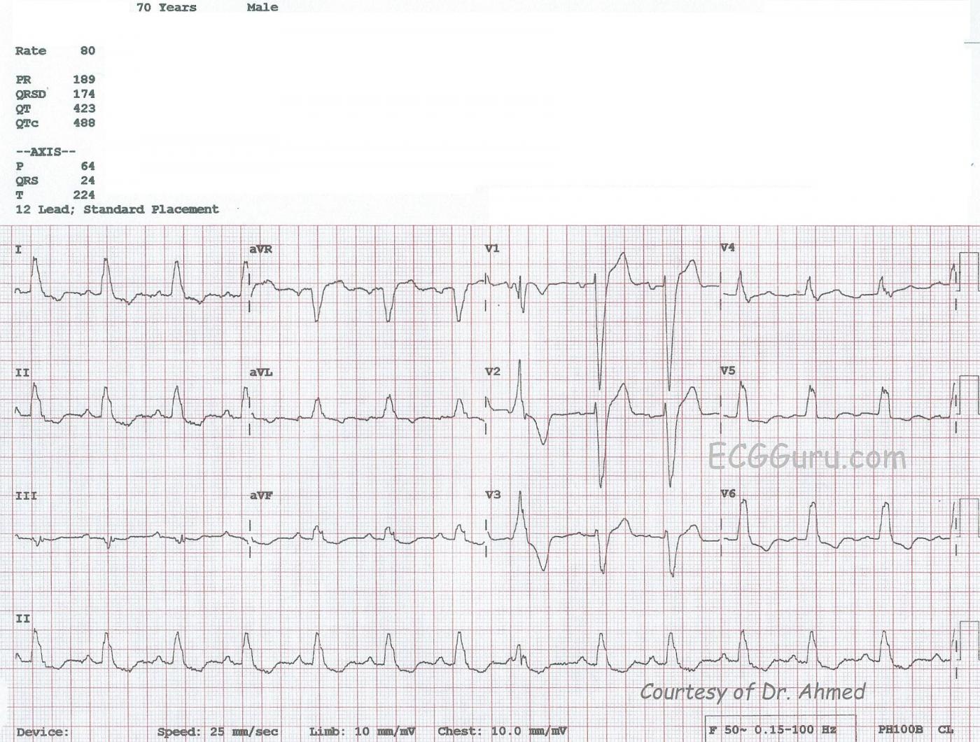
This ECG, kindly donated by Dr. Ahmed from India, is from a 70-year-old man shows a sinus rhythm at 80 bpm with left bundle branch block (LBBB), left atrial enlargement (LAE), and a premature ventricular contraction (PVC). The ECG criteria for LBBB is:  1) Wide QRS  - greater than or equal to .12 seconds;  2) Supraventricular rhythm;  3) QRS that is negative in V1 and positive in Leads I and V6. In leads with a positive QRS, we will see some ST depression, and in leads with a negative QRS, some ST elevation.  This is "normal" for the wide QRS rhythm, and does not indicate injury or ischemia, although it does not rule it out, either.  LBBB is an indicator of cardiac disease, but not specific to one etiology.

There is a PVC seen as the 8th beat from the left, and it gives you a chance to show your students a wide-complex beat that is NOT associated with a P wave and is premature, compared to the wide-complex SINUS beats with LBBB.  The PVC, being wide-complex, also has similar ST changes:  the ST segments and T waves are DISCORDANT with the QRS complexes.

The P waves show some signs of enlargement of the left atrium.  The P waves in Lead II are tall and pointed, and the P waves in V1 are biphasic.  Left atrial enlargement in a patient with LBBB would not be surprising, as both are associated with left ventricular dysfunction.  Patients with these ECG patterns should be thoroughly evaluated for congestive heart failure.  Patients with LBBB, low ejection fractions, and heart failure are treated with cardiac resynchronization therapy, using a pacemaker that paces the atria and each ventricle, synchronizing both the A-V coupling interval and the depolarization of the ventricles for optimum cardiac output.

REFERENCECirculation: Arrhythmia and Electrophysiology.  2008; 1: 127-139 doi: 10.1161/CIRCEP.108.777904

Rate this content: 
Average: 3.7 (3 votes)

Comments

ekgpress@mac.com's picture

     As per Dawn - the ECG this week illustrates complete LBBB (Left Bundle Branch Block). I'd add several points to the comments already made.
  • The 8th beat is an "end-diastolic" PVC - which means that it occurs late in the cycle. Most PVCs occur earlier the cycle. The relevance here - is that this PVC is probably a FUSION beat. Note it is preceded by a P wave with a PR interval slightly shorter than the normal PR interval for this tracing. Although impossible to prove this is a fusion beat (since we never see a "pure" PVC not preceded by a P wave) - the timing is such that I'd be surprised if there wasn't an element of fusion (ie, conduction from the preceding P wave that "fuses" in the ventricles with the PVC arising from below).
  • There are many views on what the ECG criteria for atrial enlargement ought to be. In general the ECG has poor sensitivity and poor specificity for diagnosing atrial enlargement - so I generally undercall this. My preference is for a truly deep and/or wide negative component to the P wave in lead V1. On this tracing - that criterion is barely attained. That said - given the associated LBBB - LAE becomes much more likely here. For full review on ECG criteria for left and right atrial abnormality - Please check out my ECG Blog #75.
  • There is probable LVH on this tracing. The presence of LBBB always makes it more difficult to assess for ischemia and chamber enlargement - because of the altered sequence of depolarization and repolarization. The "usual" voltage criteria for diagnosing LVH on ECG cannot be used when there is LBBB. That said - due to the fact that the overwhelming majority of patients with LBBB have significant underlying heart disease - the prevalence for finding LVH in this population is extremely high. It has been found that the likelihood of LVH is great when a patient with LBBB has either: i) very deep S waves in either lead V1,V2 or V3 that are >25-30mm; and/or ii) LAE (Left Atrial Enlargement). Both of these criteria are satisfied in this tracing - so I'd interpret this ECG as showing "probable LVH".
  • Otherwise - ST-T waves are opposite the last QRS deflection in the 3 key leads (I,V1,V6) as they should be for LBBB. There are no signs of ischemia.
For full review on the ECG diagnostic criteria for Bundle Branch Blocks - as well as assessment of associated ST-T wave changes - CLICK HERE -
 

Ken Grauer, MD  www.kg-ekgpress.com   [email protected] 

All our content is FREE & COPYRIGHT FREE for non-commercial use

Please be courteous and leave any watermark or author attribution on content you reproduce.