Dawn's picture

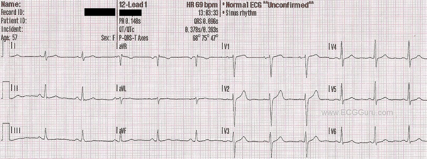
It is always good to have a number of "normal" ECGs in your collection.  This particular ECG has only three channels, lacking the fourth channel that is usually used for a rhythm strip.   Some of your students might not work in settings where they have access to four-channel ECG machines. Often, paramedics in the field and medical personnel who use "crash carts" have ECG machines with narrow paper that run three channels at once.  They will print their rhythm strips on one sheet and the 12-lead on another.  This poses a slight handicap when evaluating complex rhythms, but is adequate for most emergency ECG screenings.

This ECG has a number of features that your students should recognize as normal.  The rate and intervals are within normal ranges.  The ST segments are neither elevated or depressed.  The axis of the frontal plane leads is normal, as is the R wave progression in the chest leads.  The T waves are upright, and the P waves are uniform and upright in the inferior leads.  The rhythm is sinus.  There is no appreciable artifact.

A good understanding of what is "normal" is vital for recognizing "abnormal".

Related Terms: 
Rate this content: 
Average: 3.6 (5 votes)

Comments

ekgpress@mac.com's picture

As per Dawn - it is highly instructive to periodically mix in "normal" ECGs with abnormal ones - since one of the major tasks of the health care provider is to be comfortable distinguishing the two. This is sometimes far easier said than accomplished - especially when the patient in question is acutely symptomatic. I'd add the following points to Dawn's presentation:

  • Determining the rhythm when the mechanism is not sinus clearly becomes more challenging when one only has 3 channels available and a long lead II rhythm strip is lacking. Realize that ECGs like the one shown (in which there is no long lead II rhythm strip) are sequential - such that there are a total of 11 beats on the tracing. The 2 beats we see in lead II are enough to confirm sinus rhythm in this tracing, given that the other 9 beats are fairly regular. Slight variability in R-R interval IS allowed (the criterion for "sinus arrhythmia" is more than 0.08-to-0.12 variation in R-R interval - and that is not met here). Even when there is sinus arrhythmia - this is a common normal variant rhythm.
  • T waves are not always normally upright in all leads. While this is not an issue in this tracing - I refer you to my ECG Blog #79 for review of which leads on the normal adult ECG may manifest even deep T wave inversion or even large Q waves. Note that the T wave is upright in lead V1 of this tracing. As per Figure 2 on my ECG Blog link - the T wave in lead V1 of the normal adult ECG will often be negative, but it can be either flat, negative or upright.
  • It looks like there may be very slight J-point ST elevation in lead V2 of this tracing. The shape of this ST elevation is in an upward concavity (= "smiley"-shape) - and this is completely normal. It is good to become accustomed to slight ST elevation in anterior precordial leads in patients who you KNOW have normal ECGs - since the amount of upward concavity ST elevation that may be normally seen can be a good bit more than the tiny amount seen here.
  • S waves are present in lead I and in all 6 chest leads on this tracing. This is a descriptive finding - and not necessarily an "abnormality". Some patients with pulmonary disease manifest persist precordial S waves - whereas most normal tracings show no S wave at all in left-sided leads I, V5,V6. That said - I would not call this tracing "abnormal" because of this - but rather simply note that there are "S waves", which most probably have no clinical significance in this case.

It is often instructive for learners to see how many comments they can make about normal ECGs.

Ken Grauer, MD  www.kg-ekgpress.com   [email protected] 

All our content is FREE & COPYRIGHT FREE for non-commercial use

Please be courteous and leave any watermark or author attribution on content you reproduce.