
Submitted by Dawn on Tue, 04/02/2013 - 22:00
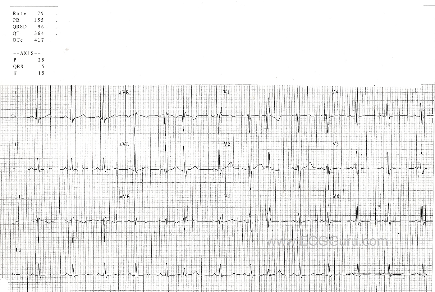
This is a normal 12-Lead ECG with two PACs that are aberrantly conducted in a right bundle branch block pattern. (Sixth and ninth beats). In the PACs, the QRS is slightly wider than the normal beats. The aberrantly-conducted beats have an rsR' pattern in V1, and a wide little S wave in aVL. No PACs are seen in Lead I to demonstrate the wide S wave. This represents a right bundle branch block pattern, which is a common form of aberrancy, and is rate-related. That is, the PAC occurs early in the cycle, catching the right bundle branch is a refractory state and unable to depolarize. Slower beats are easily acommodated by the right bundle branch.
Related Terms:
Rate this content:
All our content is FREE & COPYRIGHT FREE for non-commercial use
Please be courteous and leave any watermark or author attribution on content you reproduce.
Comments
The Basics of Aberrancy - Using 12 Leads
Perhaps the most helpful lesson learned from review of this tracing is the manner in which use of all 12-leads on an ECG may prove invaluable to arrhythmia diagnosis.
As per Dawn - there are 2 premature beats (PACs) seen in the long lead II rhythm strip at the bottom of the tracing (beats #6 and #9). That said - I would not have been certain these are PACs if the only information I had was this lead II rhythm strip. Baseline ST-T wave morphology varies somewhat among the normal sinus beats - and in lead II there is NOT a convincing premature P wave preceding beats #6 and 9. Although there is a resemblance of QRS morphology for beats #6 and 9 compared to the other beats on this tracing - a beat should always be assumed "guilty" (ie, a PVC) until proven otherwise - and there is no convincing evidence in favor of aberrant conduction in this lead II ....
On the other hand - Using the 12-lead ECG:
The above evidence from the simultaneously recorded 12-lead overwhelmingly supports a diagnosis of PACs with RBBB-aberration for early beats #6 and #9.
Ken Grauer, MD www.kg-ekgpress.com [email protected]